


 Foundational characteristics of cancer include proliferation, angiogenesis, migration, evasion of apoptosis, and cellular immortality. Find key markers for these cellular processes and antibodies to detect them.
 The SUMOplot™ Analysis Program predicts and scores sumoylation sites in your protein. SUMOylation is a post-translational modification involved in various cellular processes, such as nuclear-cytosolic transport, transcriptional regulation, apoptosis, protein stability, response to stress, and progression through the cell cycle.
 The Autophagy Receptor Motif Plotter predicts and scores autophagy receptor binding sites in your protein. Identifying proteins connected to this pathway is critical to understanding the role of autophagy in physiological as well as pathological processes such as development, differentiation, neurodegenerative diseases, stress, infection, and cancer.
Anti-HSD17B4 Picoband Antibody
- SPECIFICATION
- CITATIONS
- PROTOCOLS
- BACKGROUND

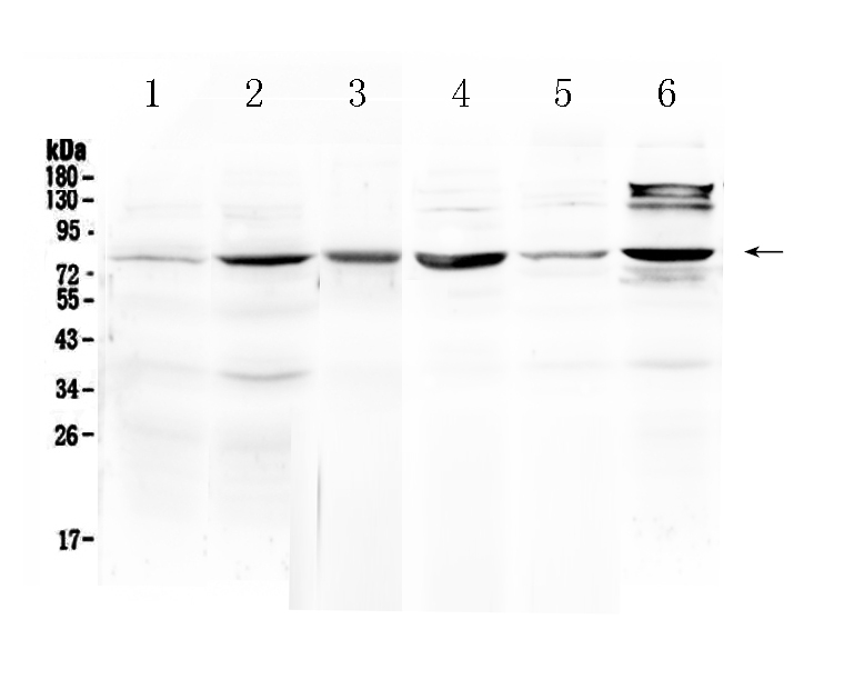
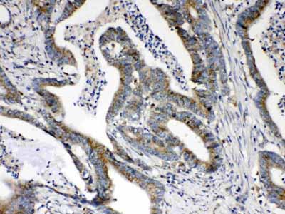
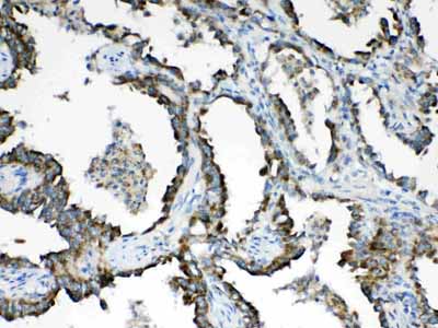
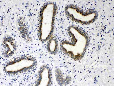
Application
| WB, IHC-P |
|---|---|
| Primary Accession | P51659 |
| Host | Rabbit |
| Reactivity | Human, Mouse, Rat |
| Clonality | Polyclonal |
| Format | Lyophilized |
| Description | Rabbit IgG polyclonal antibody for Peroxisomal multifunctional enzyme type 2(HSD17B4) detection. Tested with WB, IHC-P in Human;Mouse;Rat. |
| Reconstitution | Add 0.2ml of distilled water will yield a concentration of 500ug/ml. |
| Gene ID | 3295 |
|---|---|
| Other Names | Peroxisomal multifunctional enzyme type 2, MFE-2, 17-beta-hydroxysteroid dehydrogenase 4, 17-beta-HSD 4, D-bifunctional protein, DBP, Multifunctional protein 2, MPF-2, Short chain dehydrogenase/reductase family 8C member 1, (3R)-hydroxyacyl-CoA dehydrogenase, 1.1.1.n12, Enoyl-CoA hydratase 2, 4.2.1.107, 4.2.1.119, 3-alpha, 7-alpha, 12-alpha-trihydroxy-5-beta-cholest-24-enoyl-CoA hydratase, HSD17B4, EDH17B4, SDR8C1 |
| Calculated MW | 79686 MW KDa |
| Application Details | Immunohistochemistry(Paraffin-embedded Section), 0.5-1 µg/ml, Human, By Heat Western blot, 0.1-0.5 µg/ml, Human, Mouse, Rat |
| Subcellular Localization | Peroxisome. |
| Tissue Specificity | Present in many tissues with highest concentrations in liver, heart, prostate and testis. |
| Protein Name | Peroxisomal multifunctional enzyme type 2 |
| Contents | Each vial contains 5mg BSA, 0.9mg NaCl, 0.2mg Na2HPO4, 0.05mg NaN3. |
| Immunogen | E.coli-derived human HSD17B4 recombinant protein (Position: D510-L736). Human HSD17B4 shares 87.7% and 89% amino acid (aa) sequence identity with mouse and rat HSD17B4, respectively. |
| Purification | Immunogen affinity purified. |
| Cross Reactivity | No cross reactivity with other proteins |
| Storage | At -20˚C for one year. After r˚Constitution, at 4˚C for one month. It˚Can also be aliquotted and stored frozen at -20˚C for a longer time.Avoid repeated freezing and thawing. |
| Name | HSD17B4 (HGNC:5213) |
|---|---|
| Synonyms | EDH17B4, SDR8C1 |
| Function | Bifunctional enzyme acting on the peroxisomal fatty acid beta-oxidation pathway. Catalyzes two of the four reactions in fatty acid degradation: hydration of 2-enoyl-CoA (trans-2-enoyl-CoA) to produce (3R)-3-hydroxyacyl-CoA, and dehydrogenation of (3R)-3- hydroxyacyl-CoA to produce 3-ketoacyl-CoA (3-oxoacyl-CoA), which is further metabolized by SCPx. Can use straight-chain and branched-chain fatty acids, as well as bile acid intermediates as substrates. |
| Cellular Location | Peroxisome. |
| Tissue Location | Present in many tissues with highest concentrations in liver, heart, prostate and testis |

Thousands of laboratories across the world have published research that depended on the performance of antibodies from Abcepta to advance their research. Check out links to articles that cite our products in major peer-reviewed journals, organized by research category.
info@abcepta.com, and receive a free "I Love Antibodies" mug.

Provided below are standard protocols that you may find useful for product applications.
Background
Peroxisomal multifunctional enzyme type 2 is a protein that in humans is encoded by the HSD17B4 gene. The protein encoded by this gene is a bifunctional enzyme that is involved in the peroxisomal beta-oxidation pathway for fatty acids. It also acts as a catalyst for the formation of 3-ketoacyl-CoA intermediates from both straight-chain and 2-methyl-branched-chain fatty acids. Defects in this gene that affect the peroxisomal fatty acid beta-oxidation activity are a cause of D-bifunctional protein deficiency (DBPD). An apparent pseudogene of this gene is present on chromosome 8. Multiple alternatively spliced transcript variants encoding distinct isoforms have been found for this gene.
If you have used an Abcepta product and would like to share how it has performed, please click on the "Submit Review" button and provide the requested information. Our staff will examine and post your review and contact you if needed.
If you have any additional inquiries please email technical services at tech@abcepta.com.
Ordering Information
Other Products
Shipping Information
