


 Foundational characteristics of cancer include proliferation, angiogenesis, migration, evasion of apoptosis, and cellular immortality. Find key markers for these cellular processes and antibodies to detect them.
 The SUMOplot™ Analysis Program predicts and scores sumoylation sites in your protein. SUMOylation is a post-translational modification involved in various cellular processes, such as nuclear-cytosolic transport, transcriptional regulation, apoptosis, protein stability, response to stress, and progression through the cell cycle.
 The Autophagy Receptor Motif Plotter predicts and scores autophagy receptor binding sites in your protein. Identifying proteins connected to this pathway is critical to understanding the role of autophagy in physiological as well as pathological processes such as development, differentiation, neurodegenerative diseases, stress, infection, and cancer.
Anti-TSPAN12 Picoband Antibody
- SPECIFICATION
- CITATIONS
- PROTOCOLS
- BACKGROUND

Application
| WB, IHC-P, E |
|---|---|
| Primary Accession | O95859 |
| Host | Rabbit |
| Reactivity | Human, Mouse, Rat |
| Clonality | Polyclonal |
| Format | Lyophilized |
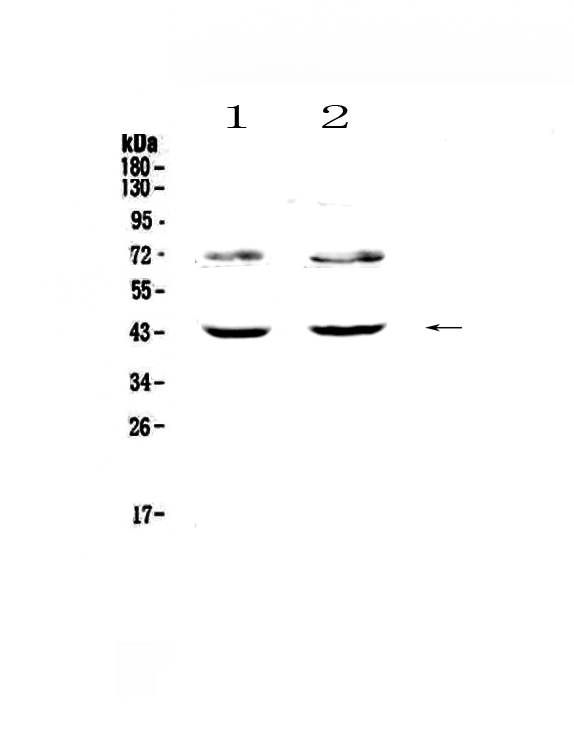
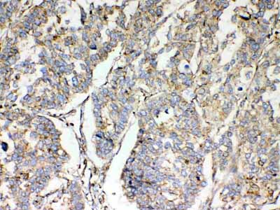
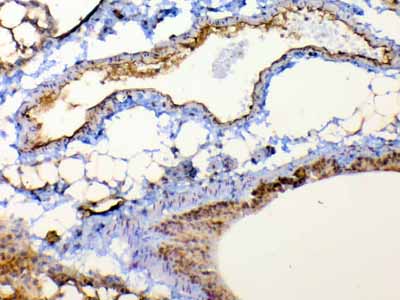
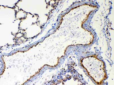
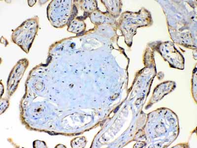
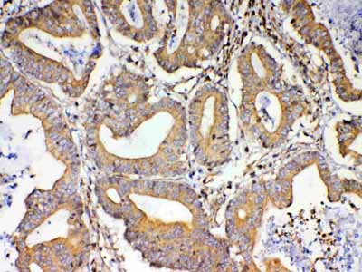
| Description | Rabbit IgG polyclonal antibody for TSPAN12 detection. Tested with WB, IHC-P, Direct ELISA in Human;Mouse;Rat. |
| Reconstitution | Add 0.2ml of distilled water will yield a concentration of 500ug/ml. |
| Gene ID | 23554 |
|---|---|
| Other Names | Tetraspanin-12, Tspan-12, Tetraspan NET-2, Transmembrane 4 superfamily member 12, TSPAN12, NET2, TM4SF12 |
| Calculated MW | 35383 Da |
| Application Details | Western blot, 0.1-0.5 µg/ml Immunohistochemistry(Paraffin-embedded Section), 0.5-1 µg/ml Direct ELISA, 0.1-0.5 µg/ml |
| Subcellular Localization | Cell membrane; Multi-pass membrane protein. |
| Contents | Each vial contains 4mg Trehalose, 0.9mg NaCl, 0.2mg Na2HPO4, 0.05mg NaN3. |
| Immunogen | E. coli-derived human TSPAN12 recombinant protein (Position: G111-R224). |
| Cross Reactivity | No cross reactivity with other proteins. |
| Storage | At -20˚C; for one year. After r˚Constitution, at 4˚C; for one month. It˚Can also be aliquotted and stored frozen at -20˚C; for a longer time. Avoid repeated freezing and thawing. |
| Name | TSPAN12 |
|---|---|
| Synonyms | NET2, TM4SF12 |
| Function | Regulator of cell surface receptor signal transduction. Plays a central role in retinal vascularization by regulating norrin (NDP) signal transduction. Acts in concert with norrin (NDP) to promote FZD4 multimerization and subsequent activation of FZD4, leading to promote accumulation of beta-catenin (CTNNB1) and stimulate LEF/TCF-mediated transcriptional programs. Suprisingly, it only activates the norrin (NDP)-dependent activation of FZD4, while it does not activate the Wnt- dependent activation of FZD4, suggesting the existence of a Wnt- independent signaling that also promote accumulation the beta-catenin (CTNNB1) (By similarity). Acts as a regulator of membrane proteinases such as ADAM10 and MMP14/MT1-MMP. Activates ADAM10-dependent cleavage activity of amyloid precursor protein (APP). Activates MMP14/MT1-MMP- dependent cleavage activity. |
| Cellular Location | Cell membrane; Multi-pass membrane protein |

Thousands of laboratories across the world have published research that depended on the performance of antibodies from Abcepta to advance their research. Check out links to articles that cite our products in major peer-reviewed journals, organized by research category.
info@abcepta.com, and receive a free "I Love Antibodies" mug.

Provided below are standard protocols that you may find useful for product applications.
Background
Tetraspanin-12 (Tspan-12) also known as tetraspan NET-2 (NET2) or transmembrane 4 superfamily member 12 (TM4SF12) is a tetraspanin protein that in humans is encoded by the TSPAN12 gene. The protein encoded by this gene is a member of the transmembrane 4 superfamily, also known as the tetraspanin family. Most of these members are cell-surface proteins that are characterized by the presence of four hydrophobic domains. The proteins mediate signal transduction events that play a role in the regulation of cell development, activation, growth and motility.
If you have used an Abcepta product and would like to share how it has performed, please click on the "Submit Review" button and provide the requested information. Our staff will examine and post your review and contact you if needed.
If you have any additional inquiries please email technical services at tech@abcepta.com.
Ordering Information
Shipping Information
