


 Foundational characteristics of cancer include proliferation, angiogenesis, migration, evasion of apoptosis, and cellular immortality. Find key markers for these cellular processes and antibodies to detect them.
 The SUMOplot™ Analysis Program predicts and scores sumoylation sites in your protein. SUMOylation is a post-translational modification involved in various cellular processes, such as nuclear-cytosolic transport, transcriptional regulation, apoptosis, protein stability, response to stress, and progression through the cell cycle.
 The Autophagy Receptor Motif Plotter predicts and scores autophagy receptor binding sites in your protein. Identifying proteins connected to this pathway is critical to understanding the role of autophagy in physiological as well as pathological processes such as development, differentiation, neurodegenerative diseases, stress, infection, and cancer.
sRANK Ligand Antibody
Rabbit Polyclonal Antibody
- SPECIFICATION
- CITATIONS
- PROTOCOLS
- BACKGROUND

Application
| WB, E |
|---|---|
| Primary Accession | O35235 |
| Reactivity | Mouse |
| Host | Rabbit |
| Clonality | Polyclonal |
| Isotype | Rabbit IgG |
| Calculated MW | 35003 Da |
| Gene ID | 21943 |
|---|---|
| Positive Control | ELISA: Recombinant msRANKL |
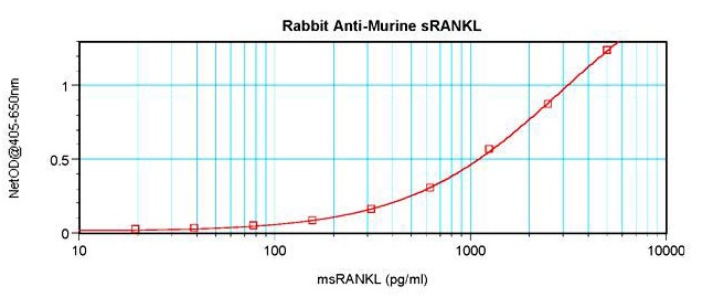
| Application & Usage | 1) WB: Use 0.1-0.2 µg/ml. The detection limit for recombinant murine sRANKL is 1.5-3.0 ng/lane, under either reducing or non-reducing conditions. 2) ELISA: Use 0.5 - 2.0 µg/ml (100 µl/well antibody solution) 3) Neutralization: To yield one-half maximal inhibition [ND50] of the biological activity of msRANKL (50.0 ng/ml), a concentration of 0.05 µg/ml of this antibody is required. |
| Other Names | soluble Receptor Activator of NFkB Ligand, TNFSF11, TRANCE (TNF-related activation-induced cytokine), OPGL, ODF (Osteoclast differentiation factor) |
| Target/Specificity | sRANKL |
| Antibody Form | Liquid |
| Appearance | Liquid |
| Formulation | A sterile filtered antibody solution in PBS, pH 7.2. |
| Handling | The antibody solution should be gently mixed before use. |
| Reconstitution & Storage | -20 °C |
| Background Descriptions | |
| Precautions | sRANK Ligand Antibody is for research use only and not for use in diagnostic or therapeutic procedures. |
| Name | Tnfsf11 |
|---|---|
| Synonyms | Opgl, Rankl, Trance |
| Function | Cytokine that binds to TNFRSF11B/OPG and to TNFRSF11A/RANK. Osteoclast differentiation and activation factor (PubMed:22437732). Augments the ability of dendritic cells to stimulate naive T-cell proliferation. May be an important regulator of interactions between T- cells and dendritic cells and may play a role in the regulation of the T-cell-dependent immune response. May also play an important role in enhanced bone-resorption in humoral hypercalcemia of malignancy (By similarity). Induces osteoclastogenesis by activating multiple signaling pathways in osteoclast precursor cells, chief among which is induction of long lasting oscillations in the intracellular concentration of Ca (2+) resulting in the activation of NFATC1, which translocates to the nucleus and induces osteoclast-specific gene transcription to allow differentiation of osteoclasts (PubMed:18586671, PubMed:24039232, PubMed:27336669). During osteoclast differentiation, in a TMEM64 and ATP2A2-dependent manner induces activation of CREB1 and mitochondrial ROS generation necessary for proper osteoclast generation (PubMed:23395171, PubMed:26644563). |
| Cellular Location | [Isoform 1]: Cell membrane; Single-pass type II membrane protein [Isoform 3]: Cytoplasm. |
| Tissue Location | Highly expressed in thymus and lymph nodes, but not in non-lymphoid tissues and is abundantly expressed in T-cells but not in B-cells. A high level expression is also seen in the trabecular bone and lung |

Thousands of laboratories across the world have published research that depended on the performance of antibodies from Abcepta to advance their research. Check out links to articles that cite our products in major peer-reviewed journals, organized by research category.
info@abcepta.com, and receive a free "I Love Antibodies" mug.

Provided below are standard protocols that you may find useful for product applications.
Background
RANKL and RANK are members of the TNF superfamily of ligands and receptors that play an important role in the regulation of specific immunity and bone turnover. RANK (receptor) was originally identified as a dendritic-cell-membrane protein, which by interacting with RANKL augments the ability of dendritic cells to stimulate naïve T-cell proliferation in a mixed lymphocyte reaction, to promote the survival of RANK + T cells, and to regulate T-cell-dependent immune response. RANKL, which is expressed in a variety of cells including osteoblasts, fibroblasts, activated T-cells and bone marrow stromal cells, is also capable of interacting with a decoy receptor called OPG. Binding of soluble OPG to sRANKL inhibits osteoclastogenesis by interrupting the signaling between stromal cells and osteoclastic progenitor cells, thereby leading to excess accumulation of bone and cartilage.
If you have used an Abcepta product and would like to share how it has performed, please click on the "Submit Review" button and provide the requested information. Our staff will examine and post your review and contact you if needed.
If you have any additional inquiries please email technical services at tech@abcepta.com.
Ordering Information
Shipping Information
