


 Foundational characteristics of cancer include proliferation, angiogenesis, migration, evasion of apoptosis, and cellular immortality. Find key markers for these cellular processes and antibodies to detect them.
 The SUMOplot™ Analysis Program predicts and scores sumoylation sites in your protein. SUMOylation is a post-translational modification involved in various cellular processes, such as nuclear-cytosolic transport, transcriptional regulation, apoptosis, protein stability, response to stress, and progression through the cell cycle.
 The Autophagy Receptor Motif Plotter predicts and scores autophagy receptor binding sites in your protein. Identifying proteins connected to this pathway is critical to understanding the role of autophagy in physiological as well as pathological processes such as development, differentiation, neurodegenerative diseases, stress, infection, and cancer.
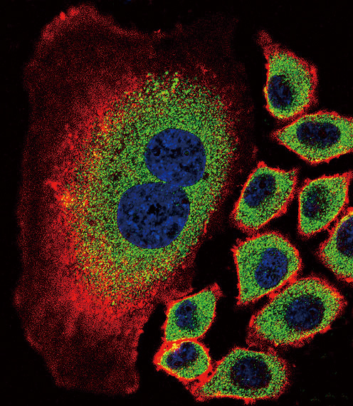
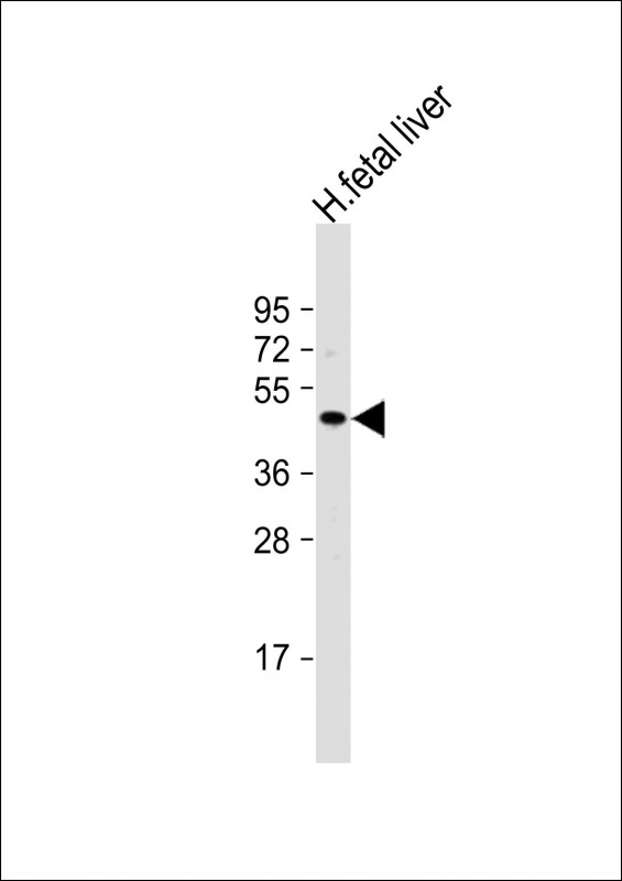
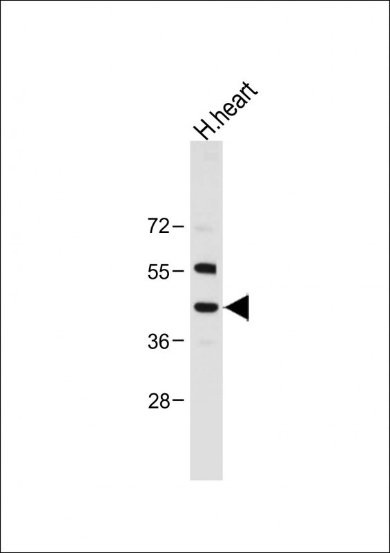
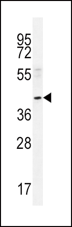
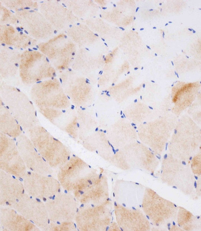
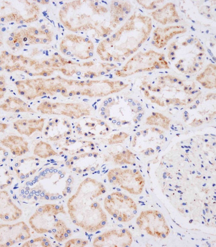
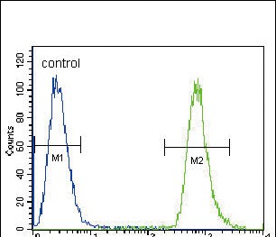
NEU2 Antibody (N-term)
Affinity Purified Rabbit Polyclonal Antibody (Pab)
- SPECIFICATION
- CITATIONS
- PROTOCOLS
- BACKGROUND

Application
| IHC-P-Leica, FC, IF, WB, E |
|---|---|
| Primary Accession | Q9Y3R4 |
| Other Accession | NP_005374.2 |
| Reactivity | Human |
| Host | Rabbit |
| Clonality | Polyclonal |
| Isotype | Rabbit IgG |
| Calculated MW | 42254 Da |
| Antigen Region | 23-50 aa |
| Gene ID | 4759 |
|---|---|
| Other Names | Sialidase-2, Cytosolic sialidase, N-acetyl-alpha-neuraminidase 2, NEU2 |
| Target/Specificity | This NEU2 antibody is generated from rabbits immunized with a KLH conjugated synthetic peptide between 23-50 amino acids from the N-terminal region of human NEU2. |
| Dilution | IF~~1:10~50 WB~~1:1000 IHC-P-Leica~~1:250 FC~~1:10~50 |
| Format | Purified polyclonal antibody supplied in PBS with 0.09% (W/V) sodium azide. This antibody is purified through a protein A column, followed by peptide affinity purification. |
| Storage | Maintain refrigerated at 2-8°C for up to 2 weeks. For long term storage store at -20°C in small aliquots to prevent freeze-thaw cycles. |
| Precautions | NEU2 Antibody (N-term) is for research use only and not for use in diagnostic or therapeutic procedures. |
| Name | NEU2 |
|---|---|
| Function | Exo-alpha-sialidase that catalyzes the hydrolytic cleavage of the terminal sialic acid (N-acetylneuraminic acid, Neu5Ac) of a glycan moiety in the catabolism of glycolipids, glycoproteins and oligosacharides (PubMed:14613940, PubMed:22228546). Recognizes sialyl linkage positions of the glycan moiety as well as the supramolecular organization of the sialoglycoconjugate. Displays preference for alpha- (2->3)-sialylated GD1a and GT1B gangliosides over alpha-(2->8)- sialylated GD1b, in both monomeric forms and micelles. Hydrolyzes monomeric GM1 ganglioside, but has no activity toward the miscellar form (PubMed:14613940). Has lower sialidase activity for glycoproteins such as fetuin and TF/transferrin that carry a mixture of alpha-(2->3) and alpha-(2->6)-sialyl linkages. Cleaves milk oligosaccharide alpha- (2->3)-sialyllactose, but is inactive toward alpha-(2->6)-sialyllactose isomer. Has no activity toward colominic acid, a homomer of alpha- (2->8)-linked Neu5Ac residues (PubMed:14613940). |
| Cellular Location | Cytoplasm, cytosol. |
| Tissue Location | Expressed in skeletal muscle, fetal liver and embryonic carcinoma cell line NT2-D1. |

Thousands of laboratories across the world have published research that depended on the performance of antibodies from Abcepta to advance their research. Check out links to articles that cite our products in major peer-reviewed journals, organized by research category.
info@abcepta.com, and receive a free "I Love Antibodies" mug.

Provided below are standard protocols that you may find useful for product applications.
Background
This gene belongs to a family of glycohydrolytic enzymes which remove sialic acid residues from glycoproteins and glycolipids. Expression studies in COS7 cells confirmed that this gene encodes a functional sialidase. Its cytosolic localization was demonstrated by cell fractionation experiments. [provided by RefSeq].
References
Stoppani, E., et al. Cell Biol. Int. 33(9):1020-1025(2009)
Li, C.Y., et al. Cell Res. 17(4):357-362(2007)
Chavas, L.M., et al. J. Biol. Chem. 280(1):469-475(2005)
Seyrantepe, V., et al. J. Biol. Chem. 279(35):37021-37029(2004)
Tringali, C., et al. J. Biol. Chem. 279(5):3169-3179(2004)
If you have used an Abcepta product and would like to share how it has performed, please click on the "Submit Review" button and provide the requested information. Our staff will examine and post your review and contact you if needed.
If you have any additional inquiries please email technical services at tech@abcepta.com.
Ordering Information
Other Products
Shipping Information
