


 Foundational characteristics of cancer include proliferation, angiogenesis, migration, evasion of apoptosis, and cellular immortality. Find key markers for these cellular processes and antibodies to detect them.
 The SUMOplot™ Analysis Program predicts and scores sumoylation sites in your protein. SUMOylation is a post-translational modification involved in various cellular processes, such as nuclear-cytosolic transport, transcriptional regulation, apoptosis, protein stability, response to stress, and progression through the cell cycle.
 The Autophagy Receptor Motif Plotter predicts and scores autophagy receptor binding sites in your protein. Identifying proteins connected to this pathway is critical to understanding the role of autophagy in physiological as well as pathological processes such as development, differentiation, neurodegenerative diseases, stress, infection, and cancer.
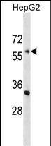
ENC1 Antibody (C-term)
Affinity Purified Rabbit Polyclonal Antibody (Pab)
- SPECIFICATION
- CITATIONS
- PROTOCOLS
- BACKGROUND

Application
| WB, E |
|---|---|
| Primary Accession | O14682 |
| Other Accession | NP_003624.1 |
| Reactivity | Human |
| Host | Rabbit |
| Clonality | Polyclonal |
| Isotype | Rabbit IgG |
| Calculated MW | 66130 Da |
| Antigen Region | 395-423 aa |
| Gene ID | 8507 |
|---|---|
| Other Names | Ectoderm-neural cortex protein 1, ENC-1, Kelch-like protein 37, Nuclear matrix protein NRP/B, p53-induced gene 10 protein, ENC1, KLHL37, NRPB, PIG10 |
| Target/Specificity | This ENC1 antibody is generated from rabbits immunized with a KLH conjugated synthetic peptide between 395-423 amino acids from the C-terminal region of human ENC1. |
| Dilution | WB~~1:1000 |
| Format | Purified polyclonal antibody supplied in PBS with 0.09% (W/V) sodium azide. This antibody is purified through a protein A column, followed by peptide affinity purification. |
| Storage | Maintain refrigerated at 2-8°C for up to 2 weeks. For long term storage store at -20°C in small aliquots to prevent freeze-thaw cycles. |
| Precautions | ENC1 Antibody (C-term) is for research use only and not for use in diagnostic or therapeutic procedures. |
| Name | ENC1 |
|---|---|
| Synonyms | KLHL37, NRPB, PIG10 |
| Function | Actin-binding protein involved in the regulation of neuronal process formation and in differentiation of neural crest cells. Down- regulates transcription factor NF2L2/NRF2 by decreasing the rate of protein synthesis and not via a ubiquitin-mediated proteasomal degradation mechanism. |
| Cellular Location | Nucleus matrix. Cytoplasm. Cytoplasm, cytoskeleton |
| Tissue Location | Detected in fetal brain tissue, moderate expression in fetal heart, lung and kidney. Highly expressed in adult brain, particularly high in the hippocampus and amygdala, and spinal chord Detectable in adult pancreas. May be down-regulated in neuroblastoma tumors |

Thousands of laboratories across the world have published research that depended on the performance of antibodies from Abcepta to advance their research. Check out links to articles that cite our products in major peer-reviewed journals, organized by research category.
info@abcepta.com, and receive a free "I Love Antibodies" mug.

Provided below are standard protocols that you may find useful for product applications.
Background
DNA damage and/or hyperproliferative signals activate wildtype p53 tumor suppressor protein (TP53; MIM 191170), inducing cell cycle arrest or apoptosis. Mutations that inactivate p53 occur in 50% of all tumors. Polyak et al. (1997) [PubMed 9305847] used serial analysis of gene expression (SAGE) to evaluate cellular mRNA levels in a colorectal cancer cell line transfected with p53. Of 7,202 transcripts identified, only 14 were expressed at levels more than 10-fold higher in p53-expressing cells than in control cells. Polyak et al. (1997) [PubMed 9305847] termed these genes 'p53-induced genes,' or PIGs, several of which were predicted to encode redox-controlling proteins. They noted that reactive oxygen species (ROS) are potent inducers of apoptosis. Flow cytometric analysis showed that p53 expression induces ROS production, which increases as apoptosis progresses under some conditions. The authors stated that the PIG10 gene, also called ENC1, encodes an actin-binding protein.
References
Seng, S., et al. Oncogene 28(3):378-389(2009)
Wang, X.J., et al. PLoS ONE 4 (5), E5492 (2009) :
Seng, S., et al. Cancer Res. 67(18):8596-8604(2007)
Barrios-Rodiles, M., et al. Science 307(5715):1621-1625(2005)
Kim, T.A., et al. Gene 255(1):105-116(2000)
If you have used an Abcepta product and would like to share how it has performed, please click on the "Submit Review" button and provide the requested information. Our staff will examine and post your review and contact you if needed.
If you have any additional inquiries please email technical services at tech@abcepta.com.

Ordering Information
Other Products
Shipping Information
