


 Foundational characteristics of cancer include proliferation, angiogenesis, migration, evasion of apoptosis, and cellular immortality. Find key markers for these cellular processes and antibodies to detect them.
 The SUMOplot™ Analysis Program predicts and scores sumoylation sites in your protein. SUMOylation is a post-translational modification involved in various cellular processes, such as nuclear-cytosolic transport, transcriptional regulation, apoptosis, protein stability, response to stress, and progression through the cell cycle.
 The Autophagy Receptor Motif Plotter predicts and scores autophagy receptor binding sites in your protein. Identifying proteins connected to this pathway is critical to understanding the role of autophagy in physiological as well as pathological processes such as development, differentiation, neurodegenerative diseases, stress, infection, and cancer.
LC3 Antibody (APG8A) (N-term)
Purified Rabbit Polyclonal Antibody (Pab)
_(N-term)_U251.jpg)
- SPECIFICATION
- CITATIONS: 28
- PROTOCOLS
- BACKGROUND

Application
| IHC-P, IF, WB, E |
|---|---|
| Primary Accession | Q9H492 |
| Reactivity | Human, Mouse, Rat |
| Host | Rabbit |
| Clonality | Polyclonal |
| Isotype | Rabbit IgG |
| Calculated MW | 14272 Da |
| Antigen Region | 1-30 aa |
| Gene ID | 84557 |
|---|---|
| Other Names | Microtubule-associated proteins 1A/1B light chain 3A, Autophagy-related protein LC3 A, Autophagy-related ubiquitin-like modifier LC3 A, MAP1 light chain 3-like protein 1, MAP1A/MAP1B light chain 3 A, MAP1A/MAP1B LC3 A, Microtubule-associated protein 1 light chain 3 alpha, MAP1LC3A |
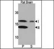
| Target/Specificity | This LC3 antibody is generated from rabbits immunized with a KLH conjugated synthetic peptide between 1~30 amino acids from the N-term of human LC3 (APG8a). |
| Dilution | IF~~1:200 WB~~1000 IHC-P~~1:50~100 |
| Format | Purified polyclonal antibody supplied in PBS with 0.09% (W/V) sodium azide. This antibody is prepared by Saturated Ammonium Sulfate (SAS) precipitation followed by dialysis against PBS. |
| Storage | Maintain refrigerated at 2-8°C for up to 2 weeks. For long term storage store at -20°C in small aliquots to prevent freeze-thaw cycles. |
| Precautions | LC3 Antibody (APG8A) (N-term) is for research use only and not for use in diagnostic or therapeutic procedures. |
| Name | MAP1LC3A (HGNC:6838) |
|---|---|
| Function | Ubiquitin-like modifier involved in formation of autophagosomal vacuoles (autophagosomes) (PubMed:20713600, PubMed:24290141). While LC3s are involved in elongation of the phagophore membrane, the GABARAP/GATE-16 subfamily is essential for a later stage in autophagosome maturation (PubMed:20713600). Through its interaction with the reticulophagy receptor TEX264, participates in the remodeling of subdomains of the endoplasmic reticulum into autophagosomes upon nutrient stress, which then fuse with lysosomes for endoplasmic reticulum turnover (PubMed:31006538, PubMed:31006537). |
| Cellular Location | Cytoplasmic vesicle, autophagosome membrane; Lipid-anchor. Endomembrane system; Lipid-anchor. Cytoplasm, cytoskeleton {ECO:0000250|UniProtKB:Q91VR7}. Note=LC3-II binds to the autophagic membranes. |
| Tissue Location | Most abundant in heart, brain, liver, skeletal muscle and testis but absent in thymus and peripheral blood leukocytes |

Provided below are standard protocols that you may find useful for product applications.
Background
Macroautophagy is the major inducible pathway for the general turnover of cytoplasmic constituents in eukaryotic cells, it is also responsible for the degradation of active cytoplasmic enzymes and organelles during nutrient starvation. Macroautophagy involves the formation of double-membrane bound autophagosomes which enclose the cytoplasmic constituent targeted for degradation in a membrane bound structure, which then fuse with the lysosome (or vacuole) releasing a single-membrane bound autophagic bodies which are then degraded within the lysosome (or vacuole). MAP1A and MAP1B are microtubule-associated proteins which mediate the physical interactions between microtubules and components of the cytoskeleton. These proteins are involved in formation of autophagosomal vacuoles (autophagosomes). MAP1A and MAP1B each consist of a heavy chain subunit and multiple light chain subunits. MAP1LC3a is one of the light chain subunits and can associate with either MAP1A or MAP1B. The precursor molecule is cleaved by APG4B/ATG4B to form the cytosolic form, LC3-I. This is activated by APG7L/ATG7, transferred to ATG3 and conjugated to phospholipid to form the membrane-bound form, LC3-II.
References
References for protein:
1.Baehrecke EH. Nat Rev Mol Cell Biol. 6(6):505-10. (2005)
2.Lum JJ, et al. Nat Rev Mol Cell Biol. 6(6):439-48. (2005)
3.Greenberg JT. Dev Cell. 8(6):799-801. (2005)
4.Levine B. Cell. 120(2):159-62. (2005)
5.Shintani T and Klionsky DJ. Science. 306(5698):990-5. (2004)
6.Tanida I., et al. Int. J. Biochem. Cell Biol. 36:2503-2518(2004)
7.He H., et al. J. Biol. Chem. 278:29278-29287(2003)
8.Tanida I., et al. J. Biol. Chem. 279:36268-36276(2004)
References for U251 cell line:
1. Westermark B.; Pontén J.; Hugosson R. (1973).” Determinants for the establishment of permanent tissue culture lines from human gliomas”. Acta Pathol Microbiol Scand A. 81:791-805. [PMID: 4359449].
2. Pontén, J.,Westermark B. (1978).” Properties of Human Malignant Glioma Cells in Vitro”. Medical Biology 56: 184-193.[PMID: 359950].
3. Geng Y.;Kohli L.; Klocke B.J.; Roth K.A.(2010). “Chloroquine-induced autophagic vacuole accumulation and cell death in glioma cells is p53 independent”. Neuro Oncol. 12(5): 473–481.[ PMID: 20406898].
If you have used an Abcepta product and would like to share how it has performed, please click on the "Submit Review" button and provide the requested information. Our staff will examine and post your review and contact you if needed.
If you have any additional inquiries please email technical services at tech@abcepta.com.
Ordering Information
Other Products
Shipping Information
