


 Foundational characteristics of cancer include proliferation, angiogenesis, migration, evasion of apoptosis, and cellular immortality. Find key markers for these cellular processes and antibodies to detect them.
 The SUMOplot™ Analysis Program predicts and scores sumoylation sites in your protein. SUMOylation is a post-translational modification involved in various cellular processes, such as nuclear-cytosolic transport, transcriptional regulation, apoptosis, protein stability, response to stress, and progression through the cell cycle.
 The Autophagy Receptor Motif Plotter predicts and scores autophagy receptor binding sites in your protein. Identifying proteins connected to this pathway is critical to understanding the role of autophagy in physiological as well as pathological processes such as development, differentiation, neurodegenerative diseases, stress, infection, and cancer.
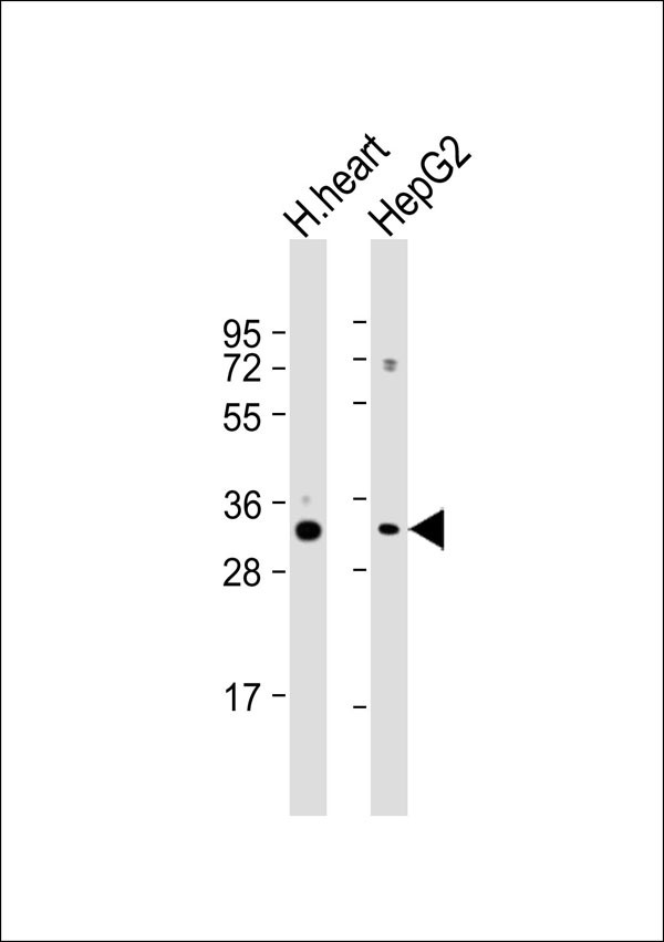
ATP5C1 Antibody
Purified Rabbit Polyclonal Antibody (Pab)
- SPECIFICATION
- CITATIONS
- PROTOCOLS
- BACKGROUND

Application
| WB |
|---|---|
| Primary Accession | P36542 |
| Reactivity | Human |
| Host | Rabbit |
| Clonality | Polyclonal |
| Calculated MW | 33 KDa |
| Antigen Region | 130-179 aa |
| Gene ID | 509 |
|---|---|
| Other Names | ATP synthase subunit gamma, mitochondrial, F-ATPase gamma subunit, ATP5C1, ATP5C, ATP5CL1 |
| Dilution | WB~~ 1:1000 |
| Format | Rabbit IgG in phosphate buffered saline , pH 7.4, 150mM NaCl, 0.09% (W/V) sodium azide and 50% glycerol |
| Storage | Store at -20 °C.Stable for 12 months from date of receipt |
| Name | ATP5F1C (HGNC:833) |
|---|---|
| Function | Mitochondrial membrane ATP synthase (F(1)F(0) ATP synthase or Complex V) produces ATP from ADP in the presence of a proton gradient across the membrane which is generated by electron transport complexes of the respiratory chain. F-type ATPases consist of two structural domains, F(1) - containing the extramembraneous catalytic core, and F(0) - containing the membrane proton channel, linked together by a central stalk and a peripheral stalk. During catalysis, ATP synthesis in the catalytic domain of F(1) is coupled via a rotary mechanism of the central stalk subunits to proton translocation. Part of the complex F(1) domain and the central stalk which is part of the complex rotary element. The gamma subunit protrudes into the catalytic domain formed of alpha(3)beta(3). Rotation of the central stalk against the surrounding alpha(3)beta(3) subunits leads to hydrolysis of ATP in three separate catalytic sites on the beta subunits. |
| Cellular Location | Mitochondrion inner membrane {ECO:0000250|UniProtKB:P05631}; Peripheral membrane protein {ECO:0000250|UniProtKB:P05631}; Matrix side {ECO:0000250|UniProtKB:P05631} |
| Tissue Location | Isoform Heart is expressed specifically in the heart and skeletal muscle, which require rapid energy supply. Isoform Liver is expressed in the brain, liver and kidney. Isoform Heart and Isoform Liver are expressed in the skin, intestine, stomach and aorta |

Thousands of laboratories across the world have published research that depended on the performance of antibodies from Abcepta to advance their research. Check out links to articles that cite our products in major peer-reviewed journals, organized by research category.
info@abcepta.com, and receive a free "I Love Antibodies" mug.

Provided below are standard protocols that you may find useful for product applications.
Background
Mitochondrial membrane ATP synthase (F(1)F(0) ATP synthase or Complex V) produces ATP from ADP in the presence of a proton gradient across the membrane which is generated by electron transport complexes of the respiratory chain. F-type ATPases consist of two structural domains, F(1) - containing the extramembraneous catalytic core, and F(0) - containing the membrane proton channel, linked together by a central stalk and a peripheral stalk. During catalysis, ATP synthesis in the catalytic domain of F(1) is coupled via a rotary mechanism of the central stalk subunits to proton translocation. Part of the complex F(1) domain and the central stalk which is part of the complex rotary element. The gamma subunit protrudes into the catalytic domain formed of alpha(3)beta(3). Rotation of the central stalk against the surrounding alpha(3)beta(3) subunits leads to hydrolysis of ATP in three separate catalytic sites on the beta subunits.
References
Matsuda C.,et al.J. Biol. Chem. 268:24950-24958(1993).
Ota T.,et al.Nat. Genet. 36:40-45(2004).
Ebert L.,et al.Submitted (JUN-2004) to the EMBL/GenBank/DDBJ databases.
Deloukas P.,et al.Nature 429:375-381(2004).
Mural R.J.,et al.Submitted (SEP-2005) to the EMBL/GenBank/DDBJ databases.
If you have used an Abcepta product and would like to share how it has performed, please click on the "Submit Review" button and provide the requested information. Our staff will examine and post your review and contact you if needed.
If you have any additional inquiries please email technical services at tech@abcepta.com.
Ordering Information
Other Products
Shipping Information
