


 Foundational characteristics of cancer include proliferation, angiogenesis, migration, evasion of apoptosis, and cellular immortality. Find key markers for these cellular processes and antibodies to detect them.
 The SUMOplot™ Analysis Program predicts and scores sumoylation sites in your protein. SUMOylation is a post-translational modification involved in various cellular processes, such as nuclear-cytosolic transport, transcriptional regulation, apoptosis, protein stability, response to stress, and progression through the cell cycle.
 The Autophagy Receptor Motif Plotter predicts and scores autophagy receptor binding sites in your protein. Identifying proteins connected to this pathway is critical to understanding the role of autophagy in physiological as well as pathological processes such as development, differentiation, neurodegenerative diseases, stress, infection, and cancer.
Anti-UNG Picoband Antibody
- SPECIFICATION
- CITATIONS
- PROTOCOLS
- BACKGROUND

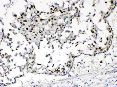
Application
| WB, IHC-P |
|---|---|
| Primary Accession | P13051 |
| Host | Rabbit |
| Reactivity | Human, Mouse, Rat |
| Clonality | Polyclonal |
| Format | Lyophilized |
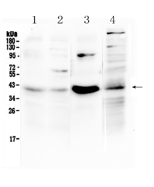
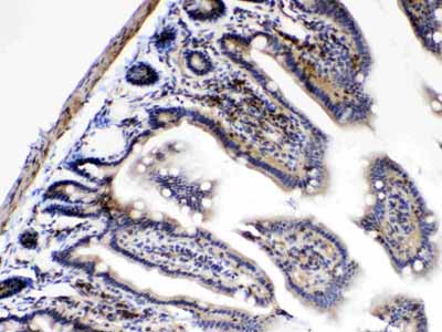
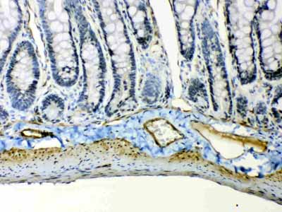
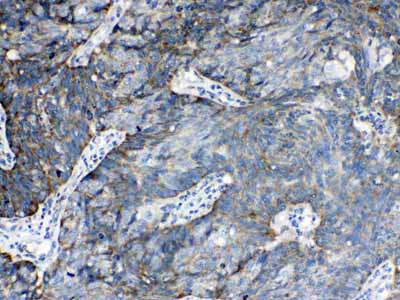
| Description | Rabbit IgG polyclonal antibody for Uracil-DNA glycosylase(UNG) detection. Tested with WB, IHC-P in Human;Mouse;Rat. |
| Reconstitution | Add 0.2ml of distilled water will yield a concentration of 500ug/ml. |
| Gene ID | 7374 |
|---|---|
| Other Names | Uracil-DNA glycosylase {ECO:0000255|HAMAP-Rule:MF_03166}, UDG {ECO:0000255|HAMAP-Rule:MF_03166}, 3.2.2.27 {ECO:0000255|HAMAP-Rule:MF_03166}, UNG {ECO:0000255|HAMAP-Rule:MF_03166} |
| Calculated MW | 34645 MW KDa |
| Application Details | Immunohistochemistry(Paraffin-embedded Section), 0.5-1 µg/ml, Human, Mouse, Rat, By Heat Western blot, 0.1-0.5 µg/ml, Human, Mouse, Rat |
| Subcellular Localization | Isoform 1: Mitochondrion. |
| Tissue Specificity | Isoform 1 is widely expressed with the highest expression in skeletal muscle, heart and testicles. Isoform 2 has the highest expression levels in tissues containing proliferating cells. |
| Protein Name | Uracil-DNA glycosylase |
| Contents | Each vial contains 5mg BSA, 0.9mg NaCl, 0.2mg Na2HPO4, 0.05mg NaN3. |
| Immunogen | E.coli-derived human UNG recombinant protein (Position: R219-L313). Human UNG shares 92.6% amino acid (aa) sequence identity with mouse UNG. |
| Purification | Immunogen affinity purified. |
| Cross Reactivity | No cross reactivity with other proteins. |
| Storage | At -20˚C for one year. After r˚Constitution, at 4˚C for one month. It˚Can also be aliquotted and stored frozen at -20˚C for a longer time.Avoid repeated freezing and thawing. |
| Name | UNG {ECO:0000255|HAMAP-Rule:MF_03166} |
|---|---|
| Function | Excises uracil residues from the DNA which can arise as a result of misincorporation of dUMP residues by DNA polymerase or due to deamination of cytosine. |
| Cellular Location | [Isoform 1]: Mitochondrion. |
| Tissue Location | Isoform 1 is widely expressed with the highest expression in skeletal muscle, heart and testicles. Isoform 2 has the highest expression levels in tissues containing proliferating cells |

Thousands of laboratories across the world have published research that depended on the performance of antibodies from Abcepta to advance their research. Check out links to articles that cite our products in major peer-reviewed journals, organized by research category.
info@abcepta.com, and receive a free "I Love Antibodies" mug.

Provided below are standard protocols that you may find useful for product applications.
Background
Uracil-DNA glycosylase, also known as UNG or UDG, is a human gene though orthologs exist ubiquitously among prokaryotes and eukaryotes and even in some DNA viruses. The first uracil DNA-glycosylase was isolated from Escherichia coli. This gene encodes one of several uracil-DNA glycosylases. One important function of uracil-DNA glycosylases is to prevent mutagenesis by eliminating uracil from DNA molecules by cleaving the N-glycosylic bond and initiating the base-excision repair (BER) pathway. Uracil bases occur from cytosine deamination or misincorporation of dUMP residues. Alternative promoter usage and splicing of this gene leads to two different isoforms: the mitochondrial UNG1 and the nuclear UNG2. The UNG2 term was used as a previous symbol for the CCNO gene, which has been confused with this gene, in the literature and some databases.
If you have used an Abcepta product and would like to share how it has performed, please click on the "Submit Review" button and provide the requested information. Our staff will examine and post your review and contact you if needed.
If you have any additional inquiries please email technical services at tech@abcepta.com.
Ordering Information
Other Products
Shipping Information
