


 Foundational characteristics of cancer include proliferation, angiogenesis, migration, evasion of apoptosis, and cellular immortality. Find key markers for these cellular processes and antibodies to detect them.
 The SUMOplot™ Analysis Program predicts and scores sumoylation sites in your protein. SUMOylation is a post-translational modification involved in various cellular processes, such as nuclear-cytosolic transport, transcriptional regulation, apoptosis, protein stability, response to stress, and progression through the cell cycle.
 The Autophagy Receptor Motif Plotter predicts and scores autophagy receptor binding sites in your protein. Identifying proteins connected to this pathway is critical to understanding the role of autophagy in physiological as well as pathological processes such as development, differentiation, neurodegenerative diseases, stress, infection, and cancer.
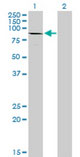
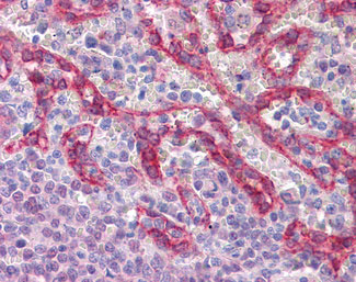
CDH1 / E Cadherin Antibody (clone 3F4)
Mouse Monoclonal Antibody
- SPECIFICATION
- CITATIONS
- PROTOCOLS
- BACKGROUND

Application
| WB, IHC-P, E, PLA |
|---|---|
| Primary Accession | P12830 |
| Reactivity | Human |
| Host | Mouse |
| Clonality | Monoclonal |
| Clone Names | 3F4 |
| Calculated MW | 97kDa |
| Dilution | IHC-P (3 µg/ml) |
| Gene ID | 999 |
|---|---|
| Other Names | Cadherin-1, CAM 120/80, Epithelial cadherin, E-cadherin, Uvomorulin, CD324, E-Cad/CTF1, E-Cad/CTF2, E-Cad/CTF3, CDH1, CDHE, UVO |
| Reconstitution & Storage | Store at -20°C. Aliquot to avoid freeze/thaw cycles. |
| Precautions | CDH1 / E Cadherin Antibody (clone 3F4) is for research use only and not for use in diagnostic or therapeutic procedures. |
| Name | CDH1 (HGNC:1748) |
|---|---|
| Function | Cadherins are calcium-dependent cell adhesion proteins (PubMed:11976333). They preferentially interact with themselves in a homophilic manner in connecting cells; cadherins may thus contribute to the sorting of heterogeneous cell types. CDH1 is involved in mechanisms regulating cell-cell adhesions, mobility and proliferation of epithelial cells (PubMed:11976333). Promotes organization of radial actin fiber structure and cellular response to contractile forces, via its interaction with AMOTL2 which facilitates anchoring of radial actin fibers to CDH1 junction complexes at the cell membrane (By similarity). Plays a role in the early stages of desmosome cell-cell junction formation via facilitating the recruitment of DSG2 and DSP to desmosome plaques (PubMed:29999492). Has a potent invasive suppressor role. It is a ligand for integrin alpha-E/beta-7. |
| Cellular Location | Cell junction, adherens junction. Cell membrane; Single-pass type I membrane protein Endosome. Golgi apparatus, trans-Golgi network. Cytoplasm. Cell junction, desmosome. Note=Colocalizes with DLGAP5 at sites of cell-cell contact in intestinal epithelial cells. Anchored to actin microfilaments through association with alpha-, beta- and gamma- catenin. Sequential proteolysis induced by apoptosis or calcium influx, results in translocation from sites of cell-cell contact to the cytoplasm. Colocalizes with RAB11A endosomes during its transport from the Golgi apparatus to the plasma membrane. Recruited to desmosomes at the initial assembly phase and also accumulates progressively at mature desmosome cell-cell junctions (PubMed:25208567, PubMed:29999492) Localizes to cell-cell contacts as keratinocyte differentiation progresses (By similarity). {ECO:0000250|UniProtKB:P09803, ECO:0000269|PubMed:25208567, ECO:0000269|PubMed:29999492} |
| Tissue Location | Expressed in granuloma macrophages (at protein level) (PubMed:27760340). Expressed in the skin (at protein level) (PubMed:22294297). Expressed in the liver (PubMed:3263290) |

Thousands of laboratories across the world have published research that depended on the performance of antibodies from Abcepta to advance their research. Check out links to articles that cite our products in major peer-reviewed journals, organized by research category.
info@abcepta.com, and receive a free "I Love Antibodies" mug.

Provided below are standard protocols that you may find useful for product applications.
Background
Cadherins are calcium-dependent cell adhesion proteins. They preferentially interact with themselves in a homophilic manner in connecting cells; cadherins may thus contribute to the sorting of heterogeneous cell types. CDH1 is involved in mechanisms regulating cell-cell adhesions, mobility and proliferation of epithelial cells. Has a potent invasive suppressor role. It is a ligand for integrin alpha-E/beta-7.
References
Bussemakers M.J.G.,et al.Mol. Biol. Rep. 17:123-128(1993).
Oda T.,et al.Proc. Natl. Acad. Sci. U.S.A. 91:1858-1862(1994).
Rimm D.L.,et al.Biochem. Biophys. Res. Commun. 200:1754-1761(1994).
Ito K.,et al.Oncogene 18:7080-7090(1999).
Shibamoto S.,et al.Submitted (MAR-1999) to the EMBL/GenBank/DDBJ databases.
If you have used an Abcepta product and would like to share how it has performed, please click on the "Submit Review" button and provide the requested information. Our staff will examine and post your review and contact you if needed.
If you have any additional inquiries please email technical services at tech@abcepta.com.
Ordering Information
Other Products
Shipping Information
