


 Foundational characteristics of cancer include proliferation, angiogenesis, migration, evasion of apoptosis, and cellular immortality. Find key markers for these cellular processes and antibodies to detect them.
 The SUMOplot™ Analysis Program predicts and scores sumoylation sites in your protein. SUMOylation is a post-translational modification involved in various cellular processes, such as nuclear-cytosolic transport, transcriptional regulation, apoptosis, protein stability, response to stress, and progression through the cell cycle.
 The Autophagy Receptor Motif Plotter predicts and scores autophagy receptor binding sites in your protein. Identifying proteins connected to this pathway is critical to understanding the role of autophagy in physiological as well as pathological processes such as development, differentiation, neurodegenerative diseases, stress, infection, and cancer.
PARD3 Antibody (aa1141-1190)
Rabbit Polyclonal Antibody
- SPECIFICATION
- CITATIONS
- PROTOCOLS
- BACKGROUND

Application
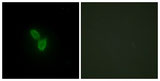
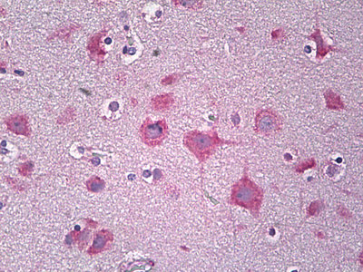
| WB, IHC-P, IF, E |
|---|---|
| Primary Accession | Q8TEW0 |
| Reactivity | Human, Mouse, Rat |
| Host | Rabbit |
| Clonality | Polyclonal |
| Calculated MW | 151kDa |
| Dilution | ELISA (1:20000), IF (1:100-1:500), IHC-P (10 µg/ml), WB (1:500-1:1000) , |
| Gene ID | 56288 |
|---|---|
| Other Names | Partitioning defective 3 homolog, PAR-3, PARD-3, Atypical PKC isotype-specific-interacting protein, ASIP, CTCL tumor antigen se2-5, PAR3-alpha, PARD3, PAR3, PAR3A |
| Target/Specificity | PARD3 Antibody detects endogenous levels of total PARD3 protein. |
| Reconstitution & Storage | Short term 4°C, long term aliquot and store at -20°C, avoid freeze thaw cycles. |
| Precautions | PARD3 Antibody (aa1141-1190) is for research use only and not for use in diagnostic or therapeutic procedures. |
| Name | PARD3 (HGNC:16051) |
|---|---|
| Synonyms | PAR3, PAR3A |
| Function | Adapter protein involved in asymmetrical cell division and cell polarization processes (PubMed:27925688, PubMed:10954424). Seems to play a central role in the formation of epithelial tight junctions (PubMed:27925688). Targets the phosphatase PTEN to cell junctions (By similarity). Involved in Schwann cell peripheral myelination (By similarity). Association with PARD6B may prevent the interaction of PARD3 with F11R/JAM1, thereby preventing tight junction assembly (By similarity). The PARD6-PARD3 complex links GTP-bound Rho small GTPases to atypical protein kinase C proteins (PubMed:10934474). Required for establishment of neuronal polarity and normal axon formation in cultured hippocampal neurons (PubMed:19812038, PubMed:27925688). |
| Cellular Location | Cytoplasm. Endomembrane system. Cell junction. Cell junction, tight junction. Cell junction, adherens junction {ECO:0000250|UniProtKB:Q99NH2}. Cell membrane. Cytoplasm, cell cortex. Cytoplasm, cytoskeleton. Note=Localized along the cell-cell contact region. Colocalizes with PARD6A and PRKCI at epithelial tight junctions. Colocalizes with the cortical actin that overlays the meiotic spindle during metaphase I and metaphase II. Colocalized with SIRT2 in internode region of myelin sheath (By similarity). Presence of KRIT1, CDH5 and RAP1B is required for its localization to the cell junction. |
| Tissue Location | Widely expressed.. |
| Volume | 50 µl |

Thousands of laboratories across the world have published research that depended on the performance of antibodies from Abcepta to advance their research. Check out links to articles that cite our products in major peer-reviewed journals, organized by research category.
info@abcepta.com, and receive a free "I Love Antibodies" mug.

Provided below are standard protocols that you may find useful for product applications.
Background
Adapter protein involved in asymmetrical cell division and cell polarization processes. Seems to play a central role in the formation of epithelial tight junctions. Targets the phosphatase PTEN to cell junctions. Involved in Schwann cell peripheral myelination (By similarity). Association with PARD6B may prevent the interaction of PARD3 with F11R/JAM1, thereby preventing tight junction assembly. The PARD6-PARD3 complex links GTP-bound Rho small GTPases to atypical protein kinase C proteins. Required for establishment of neuronal polarity and normal axon formation in cultured hippocampal neurons.
References
Joberty G.,et al.Nat. Cell Biol. 2:531-539(2000).
Fang C.M.,et al.Cell Res. 11:223-229(2001).
Kohjima M.,et al.Biochem. Biophys. Res. Commun. 299:641-646(2002).
Gao L.,et al.Gene 294:99-107(2002).
Deloukas P.,et al.Nature 429:375-381(2004).
If you have used an Abcepta product and would like to share how it has performed, please click on the "Submit Review" button and provide the requested information. Our staff will examine and post your review and contact you if needed.
If you have any additional inquiries please email technical services at tech@abcepta.com.
Ordering Information
Other Products
Shipping Information
