


 Foundational characteristics of cancer include proliferation, angiogenesis, migration, evasion of apoptosis, and cellular immortality. Find key markers for these cellular processes and antibodies to detect them.
 The SUMOplot™ Analysis Program predicts and scores sumoylation sites in your protein. SUMOylation is a post-translational modification involved in various cellular processes, such as nuclear-cytosolic transport, transcriptional regulation, apoptosis, protein stability, response to stress, and progression through the cell cycle.
 The Autophagy Receptor Motif Plotter predicts and scores autophagy receptor binding sites in your protein. Identifying proteins connected to this pathway is critical to understanding the role of autophagy in physiological as well as pathological processes such as development, differentiation, neurodegenerative diseases, stress, infection, and cancer.
Nephrin Antibody (Ascites)
Mouse Monoclonal Antibody (Mab)

- SPECIFICATION
- CITATIONS
- PROTOCOLS
- BACKGROUND

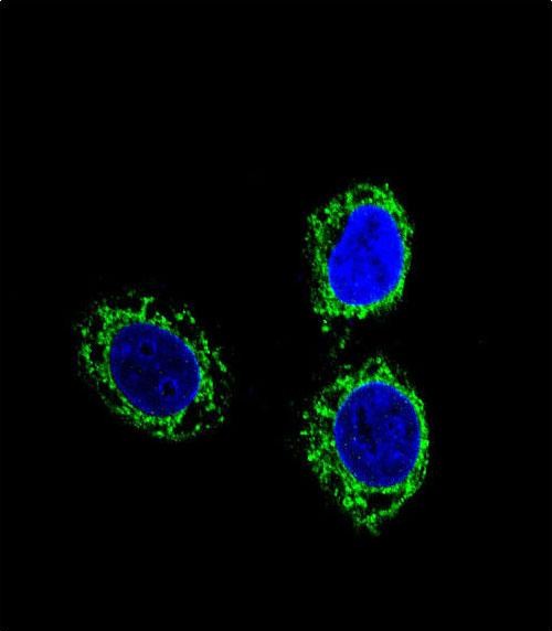
Application
| WB, IF, E |
|---|---|
| Primary Accession | O60500 |
| Other Accession | NP_004637.1 |
| Reactivity | Human |
| Host | Mouse |
| Clonality | Monoclonal |
| Isotype | IgM,K |
| Clone/Animal Names | 174CT2.1.1 |
| Calculated MW | 134742 Da |
| Antigen Region | 1088-1117 aa |
| Gene ID | 4868 |
|---|---|
| Other Names | Nephrin, Renal glomerulus-specific cell adhesion receptor, NPHS1, NPHN |
| Target/Specificity | This Nephrin antibody is generated from mice immunized with a KLH conjugated synthetic peptide between 1088-1117 amino acids from human Nephrin. |
| Dilution | WB~~1:500~1000 IF~~1:10~50 E~~Use at an assay dependent concentration. |
| Format | Purified polyclonal antibody supplied in PBS with 0.09% (W/V) sodium azide. This antibody is prepared by Saturated Ammonium Sulfate (SAS) precipitation followed by dialysis against PBS. |
| Storage | Maintain refrigerated at 2-8°C for up to 2 weeks. For long term storage store at -20°C in small aliquots to prevent freeze-thaw cycles. |
| Precautions | Nephrin Antibody (Ascites) is for research use only and not for use in diagnostic or therapeutic procedures. |
| Name | NPHS1 |
|---|---|
| Synonyms | NPHN |
| Function | Seems to play a role in the development or function of the kidney glomerular filtration barrier. Regulates glomerular vascular permeability. May anchor the podocyte slit diaphragm to the actin cytoskeleton. Plays a role in skeletal muscle formation through regulation of myoblast fusion (By similarity). |
| Cellular Location | Cell membrane; Single-pass type I membrane protein. Note=Predominantly located at podocyte slit diaphragm between podocyte foot processes. Also associated with podocyte apical plasma membrane. |
| Tissue Location | Specifically expressed in podocytes of kidney glomeruli |

Thousands of laboratories across the world have published research that depended on the performance of antibodies from Abcepta to advance their research. Check out links to articles that cite our products in major peer-reviewed journals, organized by research category.
info@abcepta.com, and receive a free "I Love Antibodies" mug.

Provided below are standard protocols that you may find useful for product applications.
Background
This gene encodes a member of the immunoglobulin family of cell adhesion molecules that functions in the glomerular filtration barrier in the kidney. The gene is primarily expressed in renal tissues, and the protein is a type-1 transmembrane protein found at the slit diaphragm of glomerular podocytes. The slit diaphragm is thought to function as an ultrafilter to exclude albumin and other plasma macromolecules in the formation of urine. Mutations in this gene result in Finnish-type congenital nephrosis 1, characterized by severe proteinuria and loss of the slit diaphragm and foot processes.
References
Bailey, S.D., et al. Diabetes Care 33(10):2250-2253(2010) Wu, F., et al. J. Am. Soc. Nephrol. 21(9):1456-1467(2010) Tossidou, I., et al. J. Biol. Chem. 285(33):25285-25295(2010) Machuca, E., et al. J. Am. Soc. Nephrol. 21(7):1209-1217(2010) Aya, K., et al. Kidney Int. 57(2):401-404(2000)
If you have used an Abcepta product and would like to share how it has performed, please click on the "Submit Review" button and provide the requested information. Our staff will examine and post your review and contact you if needed.
If you have any additional inquiries please email technical services at tech@abcepta.com.
Ordering Information
Other Products
Shipping Information
