


 Foundational characteristics of cancer include proliferation, angiogenesis, migration, evasion of apoptosis, and cellular immortality. Find key markers for these cellular processes and antibodies to detect them.
 The SUMOplot™ Analysis Program predicts and scores sumoylation sites in your protein. SUMOylation is a post-translational modification involved in various cellular processes, such as nuclear-cytosolic transport, transcriptional regulation, apoptosis, protein stability, response to stress, and progression through the cell cycle.
 The Autophagy Receptor Motif Plotter predicts and scores autophagy receptor binding sites in your protein. Identifying proteins connected to this pathway is critical to understanding the role of autophagy in physiological as well as pathological processes such as development, differentiation, neurodegenerative diseases, stress, infection, and cancer.
INA Antibody
Mouse Monoclonal Antibody (Mab)
- SPECIFICATION
- CITATIONS
- PROTOCOLS
- BACKGROUND

Application
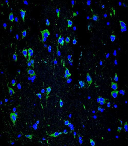
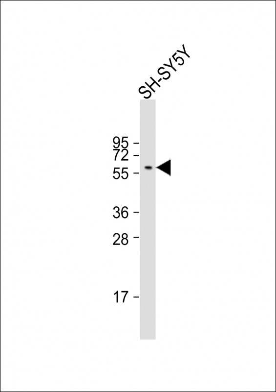
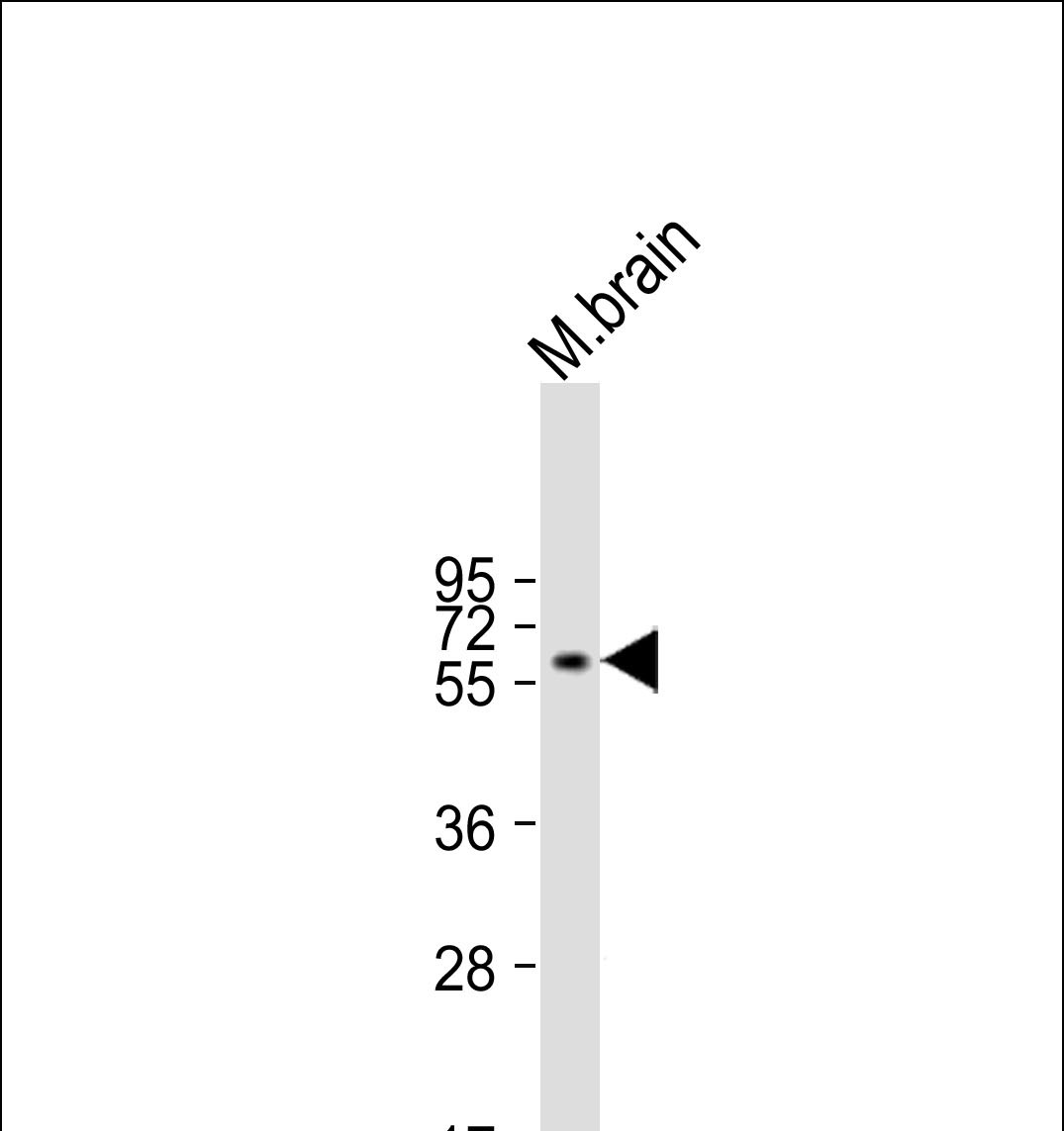
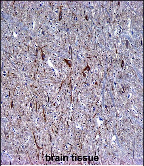
| IHC-P, IF, WB, E |
|---|---|
| Primary Accession | Q16352 |
| Other Accession | P23565, P46660, Q08DH7, NP_116116.1 |
| Reactivity | Human, Mouse |
| Predicted | Bovine, Rat |
| Host | Mouse |
| Clonality | Monoclonal |
| Isotype | IgG1,k |
| Clone/Animal Names | 257CT7.1.2 |
| Calculated MW | 55391 Da |
| Antigen Region | 290-319 aa |
| Gene ID | 9118 |
|---|---|
| Other Names | Alpha-internexin, Alpha-Inx, 66 kDa neurofilament protein, NF-66, Neurofilament-66, Neurofilament 5, INA, NEF5 |
| Target/Specificity | This INA antibody is generated from mice immunized with a KLH conjugated synthetic peptide between 290-319 amino acids from human INA. |
| Dilution | IHC-P~~1:10~50 IF~~1:10~50 WB~~1:125 E~~Use at an assay dependent concentration. |
| Format | Purified monoclonal antibody supplied in PBS with 0.09% (W/V) sodium azide. This antibody is purified through a protein G column, followed by dialysis against PBS. |
| Storage | Maintain refrigerated at 2-8°C for up to 2 weeks. For long term storage store at -20°C in small aliquots to prevent freeze-thaw cycles. |
| Precautions | INA Antibody is for research use only and not for use in diagnostic or therapeutic procedures. |
| Name | INA |
|---|---|
| Synonyms | NEF5 |
| Function | Class-IV neuronal intermediate filament that is able to self- assemble. It is involved in the morphogenesis of neurons. It may form an independent structural network without the involvement of other neurofilaments or it may cooperate with NEFL to form the filamentous backbone to which NEFM and NEFH attach to form the cross-bridges. May also cooperate with the neuronal intermediate filament protein PRPH to form filamentous networks (By similarity). |
| Tissue Location | Found predominantly in adult CNS. |

Thousands of laboratories across the world have published research that depended on the performance of antibodies from Abcepta to advance their research. Check out links to articles that cite our products in major peer-reviewed journals, organized by research category.
info@abcepta.com, and receive a free "I Love Antibodies" mug.

Provided below are standard protocols that you may find useful for product applications.
Background
Neurofilaments are type IV intermediate filament heteropolymers composed of light, medium, and heavy chains. Neurofilaments comprise the axoskeleton and they functionally maintain the neuronal caliber. They may also play a role in intracellular transport to axons and dendrites. This gene is a member of the intermediate filament family and is involved in the morphogenesis of neurons.
References
Leermakers, F.A., et al. Eur. Biophys. J. 39(9):1323-1334(2010)
Martins-de-Souza, D., et al. J Psychiatr Res 43(11):978-986(2009)
Ducray, F., et al. Neurology 72(2):156-161(2009)
Willoughby, V., et al. Appl. Immunohistochem. Mol. Morphol. 16(4):344-348(2008)
Matsuoka, S., et al. Science 316(5828):1160-1166(2007)
If you have used an Abcepta product and would like to share how it has performed, please click on the "Submit Review" button and provide the requested information. Our staff will examine and post your review and contact you if needed.
If you have any additional inquiries please email technical services at tech@abcepta.com.
Ordering Information
Other Products
Shipping Information
