


 Foundational characteristics of cancer include proliferation, angiogenesis, migration, evasion of apoptosis, and cellular immortality. Find key markers for these cellular processes and antibodies to detect them.
 The SUMOplot™ Analysis Program predicts and scores sumoylation sites in your protein. SUMOylation is a post-translational modification involved in various cellular processes, such as nuclear-cytosolic transport, transcriptional regulation, apoptosis, protein stability, response to stress, and progression through the cell cycle.
 The Autophagy Receptor Motif Plotter predicts and scores autophagy receptor binding sites in your protein. Identifying proteins connected to this pathway is critical to understanding the role of autophagy in physiological as well as pathological processes such as development, differentiation, neurodegenerative diseases, stress, infection, and cancer.
CD19 Antibody (C-term) (Ascites)
Mouse Monoclonal Antibody (Mab)
- SPECIFICATION
- CITATIONS
- PROTOCOLS
- BACKGROUND

Application
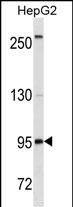
| WB, E |
|---|---|
| Primary Accession | P15391 |
| Other Accession | NP_001761.3, NP_001171569.1 |
| Reactivity | Human |
| Host | Mouse |
| Clonality | Monoclonal |
| Isotype | IgG1 |
| Clone/Animal Names | 400CT14.1.4 |
| Calculated MW | 61128 Da |
| Antigen Region | 505-532 aa |
| Gene ID | 930 |
|---|---|
| Other Names | B-lymphocyte antigen CD19, B-lymphocyte surface antigen B4, Differentiation antigen CD19, T-cell surface antigen Leu-12, CD19, CD19 |
| Target/Specificity | This CD19 antibody is generated from mice immunized with a KLH conjugated synthetic peptide between 505-532 amino acids from the C-terminal region of human CD19. |
| Dilution | WB~~1:500~1600 E~~Use at an assay dependent concentration. |
| Format | Mouse monoclonal antibody supplied in crude ascites with 0.09% (W/V) sodium azide. |
| Storage | Maintain refrigerated at 2-8°C for up to 2 weeks. For long term storage store at -20°C in small aliquots to prevent freeze-thaw cycles. |
| Precautions | CD19 Antibody (C-term) (Ascites) is for research use only and not for use in diagnostic or therapeutic procedures. |
| Name | CD19 |
|---|---|
| Function | Functions as a coreceptor for the B-cell antigen receptor complex (BCR) on B-lymphocytes (PubMed:29523808). Decreases the threshold for activation of downstream signaling pathways and for triggering B-cell responses to antigens (PubMed:1373518, PubMed:16672701, PubMed:2463100). Activates signaling pathways that lead to the activation of phosphatidylinositol 3-kinase and the mobilization of intracellular Ca(2+) stores (PubMed:12387743, PubMed:16672701, PubMed:9317126, PubMed:9382888). Is not required for early steps during B cell differentiation in the blood marrow (PubMed:9317126). Required for normal differentiation of B-1 cells (By similarity). Required for normal B cell differentiation and proliferation in response to antigen challenges (PubMed:1373518, PubMed:2463100). Required for normal levels of serum immunoglobulins, and for production of high-affinity antibodies in response to antigen challenge (PubMed:12387743, PubMed:16672701, PubMed:9317126). |
| Cellular Location | Cell membrane; Single-pass type I membrane protein. Membrane raft {ECO:0000250|UniProtKB:P25918}; Single-pass type I membrane protein {ECO:0000250|UniProtKB:P25918} |
| Tissue Location | Detected on marginal zone and germinal center B cells in lymph nodes (PubMed:2463100). Detected on blood B cells (at protein level) (PubMed:16672701, PubMed:2463100) |

Thousands of laboratories across the world have published research that depended on the performance of antibodies from Abcepta to advance their research. Check out links to articles that cite our products in major peer-reviewed journals, organized by research category.
info@abcepta.com, and receive a free "I Love Antibodies" mug.

Provided below are standard protocols that you may find useful for product applications.
Background
Lymphocytes proliferate and differentiate in response to various concentrations of different antigens. The ability of the B cell to respond in a specific, yet sensitive manner to the various antigens is achieved with the use of low-affinity antigen receptors. This gene encodes a cell surface molecule which assembles with the antigen receptor of B lymphocytes in order to decrease the threshold for antigen receptor-dependent stimulation.
References
Walter, K., et al. Oncogene 29(20):2927-2937(2010)
van Zelm, M.C., et al. J. Clin. Invest. 120(4):1265-1274(2010)
Mizuochi, T., et al. J. Interferon Cytokine Res. 30(4):243-252(2010)
Davila, S., et al. Genes Immun. 11(3):232-238(2010)
El-Sayed, Z.A., et al. Egypt J Immunol 16(1):27-38(2009)
If you have used an Abcepta product and would like to share how it has performed, please click on the "Submit Review" button and provide the requested information. Our staff will examine and post your review and contact you if needed.
If you have any additional inquiries please email technical services at tech@abcepta.com.
Ordering Information
Other Products
Shipping Information
