


 Foundational characteristics of cancer include proliferation, angiogenesis, migration, evasion of apoptosis, and cellular immortality. Find key markers for these cellular processes and antibodies to detect them.
 The SUMOplot™ Analysis Program predicts and scores sumoylation sites in your protein. SUMOylation is a post-translational modification involved in various cellular processes, such as nuclear-cytosolic transport, transcriptional regulation, apoptosis, protein stability, response to stress, and progression through the cell cycle.
 The Autophagy Receptor Motif Plotter predicts and scores autophagy receptor binding sites in your protein. Identifying proteins connected to this pathway is critical to understanding the role of autophagy in physiological as well as pathological processes such as development, differentiation, neurodegenerative diseases, stress, infection, and cancer.
VTN Antibody(Ascites)
Mouse Monoclonal Antibody (Mab)
- SPECIFICATION
- CITATIONS
- PROTOCOLS
- BACKGROUND

Application
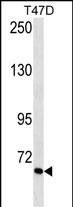
| WB, E |
|---|---|
| Primary Accession | P04004 |
| Other Accession | NP_000629.3 |
| Reactivity | Human |
| Host | Mouse |
| Clonality | Monoclonal |
| Isotype | IgM |
| Clone/Animal Names | 389CT23.2.1.3 |
| Calculated MW | 54306 Da |
| Antigen Region | 352-379 aa |
| Gene ID | 7448 |
|---|---|
| Other Names | Vitronectin, VN, S-protein, Serum-spreading factor, V75, Vitronectin V65 subunit, Vitronectin V10 subunit, Somatomedin-B, VTN |
| Target/Specificity | This VTN antibody is generated from mice immunized with a KLH conjugated synthetic peptide between 352-379 amino acids from human VTN. |
| Dilution | WB~~1:500~1600 E~~Use at an assay dependent concentration. |
| Format | Mouse monoclonal antibody supplied in crude ascites with 0.09% (W/V) sodium azide. |
| Storage | Maintain refrigerated at 2-8°C for up to 2 weeks. For long term storage store at -20°C in small aliquots to prevent freeze-thaw cycles. |
| Precautions | VTN Antibody(Ascites) is for research use only and not for use in diagnostic or therapeutic procedures. |
| Name | VTN |
|---|---|
| Function | Vitronectin is a cell adhesion and spreading factor found in serum and tissues. Vitronectin interact with glycosaminoglycans and proteoglycans. Is recognized by certain members of the integrin family and serves as a cell-to-substrate adhesion molecule. Inhibitor of the membrane-damaging effect of the terminal cytolytic complement pathway. |
| Cellular Location | Secreted, extracellular space |
| Tissue Location | Expressed in the retina pigment epithelium (at protein level) (PubMed:25136834). Expressed in plasma (at protein level) (PubMed:2448300). Expressed in serum (at protein level) (PubMed:29567995). |

Thousands of laboratories across the world have published research that depended on the performance of antibodies from Abcepta to advance their research. Check out links to articles that cite our products in major peer-reviewed journals, organized by research category.
info@abcepta.com, and receive a free "I Love Antibodies" mug.

Provided below are standard protocols that you may find useful for product applications.
Background
The protein encoded by this gene is a member of the pexin family. It is found in serum and tissues and promotes cell adhesion and spreading, inhibits the membrane-damaging effect of the terminal cytolytic complement pathway, and binds to several serpin serine protease inhibitors. It is a secreted protein and exists in either a single chain form or a clipped, two chain form held together by a disulfide bond.
References
Bailey, S.D., et al. Diabetes Care 33(10):2250-2253(2010)
Chillakuri, C.R., et al. FEBS Lett. 584(15):3287-3291(2010)
Sa E Cunha, C., et al. PLoS Pathog. 6 (5), E1000911 (2010) :
Kellouche, S., et al. Tumour Biol. 31(2):129-139(2010)
Singh, B., et al. Mol. Microbiol. 75(6):1426-1444(2010)
If you have used an Abcepta product and would like to share how it has performed, please click on the "Submit Review" button and provide the requested information. Our staff will examine and post your review and contact you if needed.
If you have any additional inquiries please email technical services at tech@abcepta.com.
Ordering Information
Other Products
Shipping Information
