


 Foundational characteristics of cancer include proliferation, angiogenesis, migration, evasion of apoptosis, and cellular immortality. Find key markers for these cellular processes and antibodies to detect them.
 The SUMOplot™ Analysis Program predicts and scores sumoylation sites in your protein. SUMOylation is a post-translational modification involved in various cellular processes, such as nuclear-cytosolic transport, transcriptional regulation, apoptosis, protein stability, response to stress, and progression through the cell cycle.
 The Autophagy Receptor Motif Plotter predicts and scores autophagy receptor binding sites in your protein. Identifying proteins connected to this pathway is critical to understanding the role of autophagy in physiological as well as pathological processes such as development, differentiation, neurodegenerative diseases, stress, infection, and cancer.
JUP Antibody (Ascites)
Mouse Monoclonal Antibody (Mab)
- SPECIFICATION
- CITATIONS
- PROTOCOLS
- BACKGROUND

Application
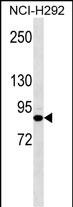
| WB, E |
|---|---|
| Primary Accession | P14923 |
| Other Accession | Q6P0K8, Q8WNW3, Q02257, Q8SPJ1, NP_002221.1 |
| Reactivity | Human |
| Predicted | Bovine, Mouse, Pig, Rat |
| Host | Mouse |
| Clonality | Monoclonal |
| Isotype | IgG3 |
| Clone/Animal Names | 606CT21.5.1 |
| Calculated MW | 81745 Da |
| Antigen Region | 636-663 aa |
| Gene ID | 3728 |
|---|---|
| Other Names | Junction plakoglobin, Catenin gamma, Desmoplakin III, Desmoplakin-3, JUP, CTNNG, DP3 |
| Target/Specificity | This JUP antibody is generated from mice immunized with a KLH conjugated synthetic peptide between 636-663 amino acids from human JUP . |
| Dilution | WB~~1:100~1600 E~~Use at an assay dependent concentration. |
| Format | Mouse monoclonal antibody supplied in crude ascites with 0.09% (W/V) sodium azide. |
| Storage | Maintain refrigerated at 2-8°C for up to 2 weeks. For long term storage store at -20°C in small aliquots to prevent freeze-thaw cycles. |
| Precautions | JUP Antibody (Ascites) is for research use only and not for use in diagnostic or therapeutic procedures. |
| Name | JUP (HGNC:6207) |
|---|---|
| Function | Common junctional plaque protein. The membrane-associated plaques are architectural elements in an important strategic position to influence the arrangement and function of both the cytoskeleton and the cells within the tissue. The presence of plakoglobin in both the desmosomes and in the intermediate junctions suggests that it plays a central role in the structure and function of submembranous plaques. Acts as a substrate for VE-PTP and is required by it to stimulate VE- cadherin function in endothelial cells. Can replace beta-catenin in E- cadherin/catenin adhesion complexes which are proposed to couple cadherins to the actin cytoskeleton (By similarity). |
| Cellular Location | Cell junction, adherens junction. Cell junction, desmosome. Cytoplasm, cytoskeleton. Cell membrane; Peripheral membrane protein. Cytoplasm {ECO:0000250|UniProtKB:Q9PVF7}. Cell junction {ECO:0000250|UniProtKB:Q9PVF7}. Nucleus {ECO:0000250|UniProtKB:Q9PVF7} Note=Cytoplasmic in a soluble and membrane-associated form. Colocalizes with DSG4 at desmosomes (PubMed:21495994) |
| Tissue Location | Expressed in the heart (at protein level). |

Thousands of laboratories across the world have published research that depended on the performance of antibodies from Abcepta to advance their research. Check out links to articles that cite our products in major peer-reviewed journals, organized by research category.
info@abcepta.com, and receive a free "I Love Antibodies" mug.

Provided below are standard protocols that you may find useful for product applications.
Background
This gene encodes a major cytoplasmic protein which is the only known constituent common to submembranous plaques of both desmosomes and intermediate junctions. This protein forms distinct complexes with cadherins and desmosomal cadherins and is a member of the catenin family since it contains a distinct repeating amino acid motif called the armadillo repeat. Mutation in this gene has been associated with Naxos disease. Alternative splicing occurs in this gene; however, not all transcripts have been fully described.
References
Fressart, V., et al. Europace 12(6):861-868(2010)
Cabral, R.M., et al. J. Invest. Dermatol. 130(6):1543-1550(2010)
Aktary, Z., et al. Oncogene 29(14):2118-2129(2010)
Pryczynicz, A., et al. Folia Histochem. Cytobiol. 48(1):128-133(2010)
Czyzewska, J., et al. Folia Histochem. Cytobiol. 48(1):37-45(2010)
If you have used an Abcepta product and would like to share how it has performed, please click on the "Submit Review" button and provide the requested information. Our staff will examine and post your review and contact you if needed.
If you have any additional inquiries please email technical services at tech@abcepta.com.
Ordering Information
Other Products
Shipping Information
