


 Foundational characteristics of cancer include proliferation, angiogenesis, migration, evasion of apoptosis, and cellular immortality. Find key markers for these cellular processes and antibodies to detect them.
 The SUMOplot™ Analysis Program predicts and scores sumoylation sites in your protein. SUMOylation is a post-translational modification involved in various cellular processes, such as nuclear-cytosolic transport, transcriptional regulation, apoptosis, protein stability, response to stress, and progression through the cell cycle.
 The Autophagy Receptor Motif Plotter predicts and scores autophagy receptor binding sites in your protein. Identifying proteins connected to this pathway is critical to understanding the role of autophagy in physiological as well as pathological processes such as development, differentiation, neurodegenerative diseases, stress, infection, and cancer.
CD33 Antibody
Mouse Monoclonal Antibody (Mab)
- SPECIFICATION
- CITATIONS
- PROTOCOLS
- BACKGROUND

Application
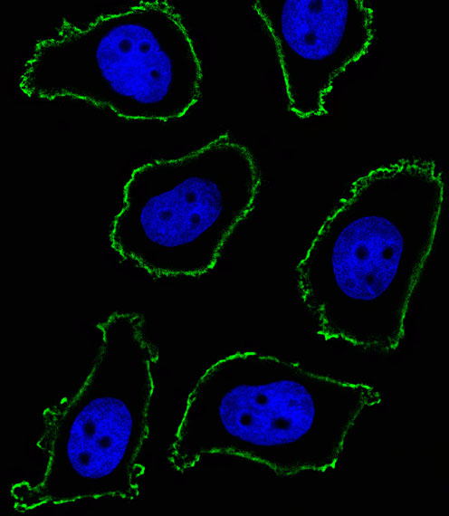
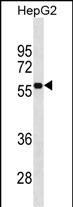
| IF, WB, E |
|---|---|
| Primary Accession | P20138 |
| Other Accession | NP_001763.3 |
| Reactivity | Human |
| Host | Mouse |
| Clonality | Monoclonal |
| Isotype | IgM |
| Clone/Animal Names | 574CT16.4.4 |
| Calculated MW | 39825 Da |
| Antigen Region | 337-364 aa |
| Gene ID | 945 |
|---|---|
| Other Names | Myeloid cell surface antigen CD33, Sialic acid-binding Ig-like lectin 3, Siglec-3, gp67, CD33, CD33, SIGLEC3 |
| Target/Specificity | This CD33 antibody is generated from mice immunized with a KLH conjugated synthetic peptide between 337-364 amino acids from human CD33. |
| Dilution | IF~~1:10~50 WB~~1:500~1000 E~~Use at an assay dependent concentration. |
| Format | Purified monoclonal antibody supplied in PBS with 0.09% (W/V) sodium azide. This antibody is prepared by Euglobin precipitation followed by dialysis against PBS. |
| Storage | Maintain refrigerated at 2-8°C for up to 2 weeks. For long term storage store at -20°C in small aliquots to prevent freeze-thaw cycles. |
| Precautions | CD33 Antibody is for research use only and not for use in diagnostic or therapeutic procedures. |
| Name | CD33 |
|---|---|
| Synonyms | SIGLEC3 |
| Function | Sialic-acid-binding immunoglobulin-like lectin (Siglec) that plays a role in mediating cell-cell interactions and in maintaining immune cells in a resting state (PubMed:10611343, PubMed:11320212, PubMed:15597323). Preferentially recognizes and binds alpha-2,3- and more avidly alpha-2,6-linked sialic acid-bearing glycans (PubMed:7718872). Upon engagement of ligands such as C1q or syalylated glycoproteins, two immunoreceptor tyrosine-based inhibitory motifs (ITIMs) located in CD33 cytoplasmic tail are phosphorylated by Src-like kinases such as LCK (PubMed:10887109, PubMed:28325905). These phosphorylations provide docking sites for the recruitment and activation of protein-tyrosine phosphatases PTPN6/SHP-1 and PTPN11/SHP- 2 (PubMed:10206955, PubMed:10556798, PubMed:10887109). In turn, these phosphatases regulate downstream pathways through dephosphorylation of signaling molecules (PubMed:10206955, PubMed:10887109). One of the repressive effect of CD33 on monocyte activation requires phosphoinositide 3-kinase/PI3K (PubMed:15597323). |
| Cellular Location | [Isoform CD33M]: Cell membrane; Single-pass type I membrane protein |
| Tissue Location | Monocytic/myeloid lineage cells. In the brain, CD33 is mainly expressed on microglial cells |

Thousands of laboratories across the world have published research that depended on the performance of antibodies from Abcepta to advance their research. Check out links to articles that cite our products in major peer-reviewed journals, organized by research category.
info@abcepta.com, and receive a free "I Love Antibodies" mug.

Provided below are standard protocols that you may find useful for product applications.
Background
Putative adhesion molecule of myelomonocytic-derived cells that mediates sialic-acid dependent binding to cells. Preferentially binds to alpha-2,6-linked sialic acid. The sialic acid recognition site may be masked by cis interactions with sialic acids on the same cell surface. In the immune response, may act as an inhibitory receptor upon ligand induced tyrosine phosphorylation by recruiting cytoplasmic phosphatase(s) via their SH2 domain(s) that block signal transduction through dephosphorylation of signaling molecules. Induces apoptosis in acute myeloid leukemia (in vitro).
References
Rose, J.E., et al. Mol. Med. 16 (7-8), 247-253 (2010) :
Davila, S., et al. Genes Immun. 11(3):232-238(2010)
Akahane, K., et al. Leukemia 24(4):865-869(2010)
Shamsasenjan, K., et al. Int. J. Hematol. 89(3):310-318(2009)
Bertram, L., et al. Am. J. Hum. Genet. 83(5):623-632(2008)
If you have used an Abcepta product and would like to share how it has performed, please click on the "Submit Review" button and provide the requested information. Our staff will examine and post your review and contact you if needed.
If you have any additional inquiries please email technical services at tech@abcepta.com.
Ordering Information
Other Products
Shipping Information
