


 Foundational characteristics of cancer include proliferation, angiogenesis, migration, evasion of apoptosis, and cellular immortality. Find key markers for these cellular processes and antibodies to detect them.
 The SUMOplot™ Analysis Program predicts and scores sumoylation sites in your protein. SUMOylation is a post-translational modification involved in various cellular processes, such as nuclear-cytosolic transport, transcriptional regulation, apoptosis, protein stability, response to stress, and progression through the cell cycle.
 The Autophagy Receptor Motif Plotter predicts and scores autophagy receptor binding sites in your protein. Identifying proteins connected to this pathway is critical to understanding the role of autophagy in physiological as well as pathological processes such as development, differentiation, neurodegenerative diseases, stress, infection, and cancer.
HAGH Antibody (C-term) (Ascites)
Mouse Monoclonal Antibody (Mab)
- SPECIFICATION
- CITATIONS
- PROTOCOLS
- BACKGROUND

Application
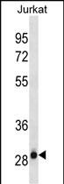
| WB, E |
|---|---|
| Primary Accession | Q16775 |
| Other Accession | NP_005317.2 |
| Reactivity | Human |
| Host | Mouse |
| Clonality | Monoclonal |
| Isotype | IgG1 |
| Clone/Animal Names | 611CT23.6.1 |
| Calculated MW | 33806 Da |
| Antigen Region | 279-308 aa |
| Gene ID | 3029 |
|---|---|
| Other Names | Hydroxyacylglutathione hydrolase, mitochondrial, Glyoxalase II, Glx II, HAGH, GLO2, HAGH1 |
| Target/Specificity | This HAGH antibody is generated from mice immunized with a KLH conjugated synthetic peptide between 279-308 amino acids from the C-terminal region of human HAGH. |
| Dilution | WB~~1:100~1600 E~~Use at an assay dependent concentration. |
| Format | Mouse monoclonal antibody supplied in crude ascites with 0.09% (W/V) sodium azide. |
| Storage | Maintain refrigerated at 2-8°C for up to 2 weeks. For long term storage store at -20°C in small aliquots to prevent freeze-thaw cycles. |
| Precautions | HAGH Antibody (C-term) (Ascites) is for research use only and not for use in diagnostic or therapeutic procedures. |
| Name | HAGH |
|---|---|
| Synonyms | GLO2, HAGH1 |
| Function | Thiolesterase that catalyzes the hydrolysis of S-D-lactoyl- glutathione to form glutathione and D-lactic acid. |
| Cellular Location | [Isoform 1]: Mitochondrion matrix |
| Tissue Location | Expressed in liver and kidney. |

Thousands of laboratories across the world have published research that depended on the performance of antibodies from Abcepta to advance their research. Check out links to articles that cite our products in major peer-reviewed journals, organized by research category.
info@abcepta.com, and receive a free "I Love Antibodies" mug.

Provided below are standard protocols that you may find useful for product applications.
Background
The enzyme encoded by this gene is classified as a thiolesterase and is responsible for the hydrolysis of S-lactoyl-glutathione to reduced glutathione and D-lactate. Two transcript variants encoding different isoforms have been found for this gene.
References
Davila, S., et al. Genes Immun. 11(3):232-238(2010)
Limphong, P., et al. Biochemistry 48(23):5426-5434(2009)
Antognelli, C., et al. Cancer Biol. Ther. 6(12):1880-1888(2007)
Xu, Y., et al. J. Biol. Chem. 281(36):26702-26713(2006)
Antognelli, C., et al. Cancer J 12(3):222-228(2006)
If you have used an Abcepta product and would like to share how it has performed, please click on the "Submit Review" button and provide the requested information. Our staff will examine and post your review and contact you if needed.
If you have any additional inquiries please email technical services at tech@abcepta.com.
Ordering Information
Other Products
Shipping Information
