


 Foundational characteristics of cancer include proliferation, angiogenesis, migration, evasion of apoptosis, and cellular immortality. Find key markers for these cellular processes and antibodies to detect them.
 The SUMOplot™ Analysis Program predicts and scores sumoylation sites in your protein. SUMOylation is a post-translational modification involved in various cellular processes, such as nuclear-cytosolic transport, transcriptional regulation, apoptosis, protein stability, response to stress, and progression through the cell cycle.
 The Autophagy Receptor Motif Plotter predicts and scores autophagy receptor binding sites in your protein. Identifying proteins connected to this pathway is critical to understanding the role of autophagy in physiological as well as pathological processes such as development, differentiation, neurodegenerative diseases, stress, infection, and cancer.
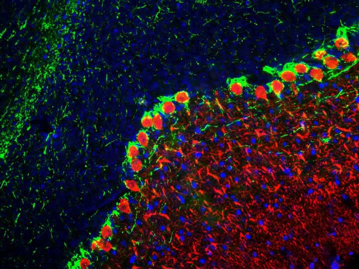
Calbindin Antibody
Mouse Monoclonal Antibody

- SPECIFICATION
- CITATIONS
- PROTOCOLS
- BACKGROUND

Application
| WB, IHC, IF |
|---|---|
| Primary Accession | P05937 |
| Reactivity | Human, Mouse, Bovine |
| Host | Mouse |
| Clonality | Monoclonal |
| Isotype | IgG2a |
| Calculated MW | 30025 Da |
| Gene ID | 793 |
|---|---|
| Gene Name | CALB1 |
| Target/Specificity | Recombinant full length human calbindin purified from E. coli. |
| Dilution | WB~~ 1:1000 IHC~~1:100~500 IF~~ 1:1000 |
| Format | Protein G purified culture supernatant |
| Storage | Maintain refrigerated at 2-8°C for up to 6 months. For long term storage store at -20°C in small aliquots to prevent freeze-thaw cycles. |
| Precautions | Calbindin Antibody is for research use only and not for use in diagnostic or therapeutic procedures. |
| Shipping | Blue Ice |

Thousands of laboratories across the world have published research that depended on the performance of antibodies from Abcepta to advance their research. Check out links to articles that cite our products in major peer-reviewed journals, organized by research category.
info@abcepta.com, and receive a free "I Love Antibodies" mug.

Provided below are standard protocols that you may find useful for product applications.
Background
Calbindin (aka. calbindin D28k) is a member of the calcium-binding protein superfamily that includes calmodulin and troponin C. It is predominantly expressed in certain types of neurons, particularly in dendrites and perikarya of the cerebellum and is thought to buffer entry of calcium upon stimulation of glutamate receptors (Andressen et al., 1993). Calbindin has recently been shown to play a critical role in mitochondrial dysfunction and loss of synaptic proteins in vivo in an Alzheimer’s disease mouse model (Kook et al., 2014).
If you have used an Abcepta product and would like to share how it has performed, please click on the "Submit Review" button and provide the requested information. Our staff will examine and post your review and contact you if needed.
If you have any additional inquiries please email technical services at tech@abcepta.com.
Ordering Information
Other Products
Shipping Information
