


 Foundational characteristics of cancer include proliferation, angiogenesis, migration, evasion of apoptosis, and cellular immortality. Find key markers for these cellular processes and antibodies to detect them.
 The SUMOplot™ Analysis Program predicts and scores sumoylation sites in your protein. SUMOylation is a post-translational modification involved in various cellular processes, such as nuclear-cytosolic transport, transcriptional regulation, apoptosis, protein stability, response to stress, and progression through the cell cycle.
 The Autophagy Receptor Motif Plotter predicts and scores autophagy receptor binding sites in your protein. Identifying proteins connected to this pathway is critical to understanding the role of autophagy in physiological as well as pathological processes such as development, differentiation, neurodegenerative diseases, stress, infection, and cancer.
MYCT1 Antibody (C-term)
Affinity Purified Rabbit Polyclonal Antibody (Pab)
- SPECIFICATION
- CITATIONS
- PROTOCOLS
- BACKGROUND

Application
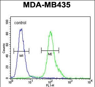
| FC, WB, E |
|---|---|
| Primary Accession | Q8N699 |
| Other Accession | NP_079383.2 |
| Reactivity | Human |
| Host | Rabbit |
| Clonality | Polyclonal |
| Isotype | Rabbit IgG |
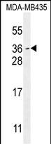
| Calculated MW | 26593 Da |
| Antigen Region | 135-163 aa |
| Gene ID | 80177 |
|---|---|
| Other Names | Myc target protein 1, Myc target in myeloid cells protein 1, MYCT1, MTLC, MTMC1 |
| Target/Specificity | This MYCT1 antibody is generated from rabbits immunized with a KLH conjugated synthetic peptide between 135-163 amino acids from the C-terminal region of human MYCT1. |
| Dilution | FC~~1:10~50 WB~~1:1000 E~~Use at an assay dependent concentration. |
| Format | Purified polyclonal antibody supplied in PBS with 0.09% (W/V) sodium azide. This antibody is purified through a protein A column, followed by peptide affinity purification. |
| Storage | Maintain refrigerated at 2-8°C for up to 2 weeks. For long term storage store at -20°C in small aliquots to prevent freeze-thaw cycles. |
| Precautions | MYCT1 Antibody (C-term) is for research use only and not for use in diagnostic or therapeutic procedures. |
| Name | MYCT1 |
|---|---|
| Synonyms | MTLC, MTMC1 |
| Function | May regulate certain MYC target genes, MYC seems to be a direct upstream transcriptional activator. Does not seem to significantly affect growth cell capacity. Overexpression seems to mediate many of the known phenotypic features associated with MYC, including promotion of apoptosis, alteration of morphology, enhancement of anchorage-independent growth, tumorigenic conversion, promotion of genomic instability, and inhibition of hematopoietic differentiation (By similarity). |
| Cellular Location | Nucleus. Note=Expressed in nuclei of hepatocellular carcinoma cell line BEL-7402 cells |
| Tissue Location | Down-regulated in gastric cancer tissues. |

Thousands of laboratories across the world have published research that depended on the performance of antibodies from Abcepta to advance their research. Check out links to articles that cite our products in major peer-reviewed journals, organized by research category.
info@abcepta.com, and receive a free "I Love Antibodies" mug.

Provided below are standard protocols that you may find useful for product applications.
Background
May regulate certain MYC target genes, MYC seems to be a direct upstream transcriptional activator. Does not seem to significantly affect growth cell capacity. Overexpression seems to mediate many of the known phenotypic features associated with MYC, including promotion of apoptosis, alteration of morphology, enhancement of anchorage-independent growth, tumorigenic conversion, promotion of genomic instability, and inhibition of hematopoietic differentiation (By similarity).
References
Qiu, G.B., et al. World J. Gastroenterol. 9(10):2160-2163(2003)
Qiu, G., et al. Zhonghua Yi Xue Yi Chuan Xue Za Zhi 20(2):94-97(2003)
If you have used an Abcepta product and would like to share how it has performed, please click on the "Submit Review" button and provide the requested information. Our staff will examine and post your review and contact you if needed.
If you have any additional inquiries please email technical services at tech@abcepta.com.
Ordering Information
Other Products
Shipping Information
