


 Foundational characteristics of cancer include proliferation, angiogenesis, migration, evasion of apoptosis, and cellular immortality. Find key markers for these cellular processes and antibodies to detect them.
 The SUMOplot™ Analysis Program predicts and scores sumoylation sites in your protein. SUMOylation is a post-translational modification involved in various cellular processes, such as nuclear-cytosolic transport, transcriptional regulation, apoptosis, protein stability, response to stress, and progression through the cell cycle.
 The Autophagy Receptor Motif Plotter predicts and scores autophagy receptor binding sites in your protein. Identifying proteins connected to this pathway is critical to understanding the role of autophagy in physiological as well as pathological processes such as development, differentiation, neurodegenerative diseases, stress, infection, and cancer.
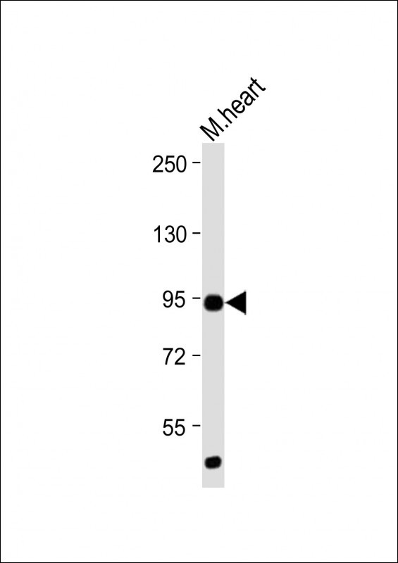
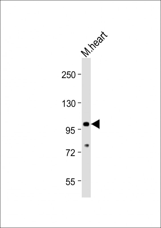
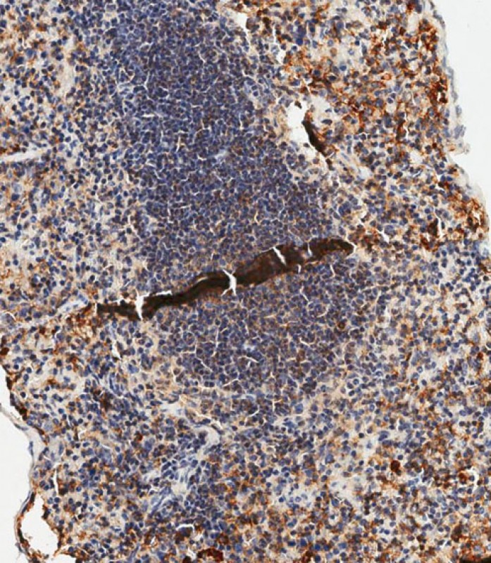
Mouse TLR2 Antibody (C-term)
Purified Rabbit Polyclonal Antibody (Pab)
- SPECIFICATION
- CITATIONS: 1
- PROTOCOLS
- BACKGROUND

Application
| WB, IHC-P-Leica, E |
|---|---|
| Primary Accession | Q9QUN7 |
| Reactivity | Mouse |
| Host | Rabbit |
| Clonality | Polyclonal |
| Isotype | Rabbit IgG |
| Calculated MW | 89449 Da |
| Antigen Region | 720-749 aa |
| Gene ID | 24088 |
|---|---|
| Other Names | Toll-like receptor 2, CD282, Tlr2 |
| Target/Specificity | This Mouse TLR2 antibody is generated from rabbits immunized with a KLH conjugated synthetic peptide between 720-749 amino acids from the C-terminal region of mouse TLR2. |
| Dilution | WB~~1:2000 IHC-P-Leica~~1:500 |
| Format | Purified polyclonal antibody supplied in PBS with 0.09% (W/V) sodium azide. This antibody is purified through a protein A column, followed by peptide affinity purification. |
| Storage | Maintain refrigerated at 2-8°C for up to 2 weeks. For long term storage store at -20°C in small aliquots to prevent freeze-thaw cycles. |
| Precautions | Mouse TLR2 Antibody (C-term) is for research use only and not for use in diagnostic or therapeutic procedures. |
| Name | Tlr2 |
|---|---|
| Function | Cooperates with LY96 to mediate the innate immune response to bacterial lipoproteins and other microbial cell wall components. Cooperates with TLR1 or TLR6 to mediate the innate immune response to bacterial lipoproteins or lipopeptides. Acts via MYD88 and TRAF6, leading to NF-kappa-B activation, cytokine secretion and the inflammatory response (By similarity) (PubMed:15690042). May also promote apoptosis in response to lipoproteins (By similarity). Forms activation clusters composed of several receptors depending on the ligand, these clusters trigger signaling from the cell surface and subsequently are targeted to the Golgi in a lipid-raft dependent pathway. Forms the cluster TLR2:TLR6:CD14:CD36 in response to diacylated lipopeptides and TLR2:TLR1:CD14 in response to triacylated lipopeptides (By similarity). Recognizes M.tuberculosis major T-antigen EsxA (ESAT-6) which inhibits downstream MYD88-dependent signaling (PubMed:17486091). Acts as the major receptor for M.tuberculosis lipoproteins LprA, LprG, LpqH and PhoS1 (pstS1), in conjunction with TLR1 and for some but not all lipoproteins CD14 and/or CD36. The lipoproteins act as agonists to modulate antigen presenting cell functions in response to the pathogen (PubMed:19362712). Recombinant MPT83 from M.tuberculosis stimulates secretion of cytokines (TNF-alpha, IL-6 and IL-12p40) by mouse macrophage cell lines in a TLR2-dependent fashion, which leads to increased host innate immunity responses against the bacterium (PubMed:22174456). Lung macrophages which express low levels of TLR2 respond poorly to stimulation by M.tuberculosis LpqH (PubMed:19362712). Required for normal uptake of M.tuberculosis, a process that is inhibited by M.tuberculosis LppM (PubMed:27220037). Interacts with TICAM2 (By similarity). |
| Cellular Location | Cell membrane; Single-pass type I membrane protein. Cytoplasmic vesicle, phagosome membrane; Single-pass type I membrane protein. Membrane raft {ECO:0000250|UniProtKB:O60603}. Note=Does not reside in lipid rafts before stimulation but accumulates increasingly in the raft upon the presence of the microbial ligand. In response to diacylated lipoproteins, TLR2:TLR6 heterodimers are recruited in lipid rafts, this recruitment determine the intracellular targeting to the Golgi apparatus. Triacylated lipoproteins induce the same mechanism for TLR2:TLR1 heterodimers. {ECO:0000250|UniProtKB:O60603} |
| Tissue Location | Detected in a macrophage cell line, smooth muscle, lung, spleen, thymus, brain and adipose tissue. Cell surface expression detected in lung alveolar macrophages, dendritic macrophages and at lower levels in lung macrophages (at protein level) (PubMed:19362712) |

Provided below are standard protocols that you may find useful for product applications.
Background
TLR2 is a member of the Toll-like receptor (TLR) family which plays a fundamental role in pathogen recognition and activation of innate immunity. TLRs are highly conserved from Drosophila to humans and share structural and functional similarities. They recognize pathogen-associated molecular patterns (PAMPs) that are expressed on infectious agents, and mediate the production of cytokines necessary for the development of effective immunity. The various TLRs exhibit different patterns of expression. TLR2 is expressed most abundantly in peripheral blood leukocytes, and mediates host response to Gram-positive bacteria and yeast via stimulation of NF-kappaB.
References
Okazaki, Y., et al., Nature 420(6915):563-573 (2002). Lin, Y., et al., J. Biol. Chem. 275(32):24255-24263 (2000). Matsuguchi, T., et al., Blood 95(4):1378-1385 (2000). Heine, H., et al., J. Immunol. 162(12):6971-6975 (1999). Underhill, D.M., et al., Nature 401(6755):811-815 (1999).
If you have used an Abcepta product and would like to share how it has performed, please click on the "Submit Review" button and provide the requested information. Our staff will examine and post your review and contact you if needed.
If you have any additional inquiries please email technical services at tech@abcepta.com.
Ordering Information
Shipping Information
