


 Foundational characteristics of cancer include proliferation, angiogenesis, migration, evasion of apoptosis, and cellular immortality. Find key markers for these cellular processes and antibodies to detect them.
 The SUMOplot™ Analysis Program predicts and scores sumoylation sites in your protein. SUMOylation is a post-translational modification involved in various cellular processes, such as nuclear-cytosolic transport, transcriptional regulation, apoptosis, protein stability, response to stress, and progression through the cell cycle.
 The Autophagy Receptor Motif Plotter predicts and scores autophagy receptor binding sites in your protein. Identifying proteins connected to this pathway is critical to understanding the role of autophagy in physiological as well as pathological processes such as development, differentiation, neurodegenerative diseases, stress, infection, and cancer.
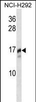
TRAPPC2 Antibody (N-term)
Affinity Purified Rabbit Polyclonal Antibody (Pab)
- SPECIFICATION
- CITATIONS
- PROTOCOLS
- BACKGROUND

Application
| WB, E |
|---|---|
| Primary Accession | P0DI81 |
| Other Accession | F1SRI0, Q9CQP2, Q08CN0, Q5ZKP4, Q3T0F2, P0DI82 |
| Reactivity | Human |
| Predicted | Bovine, Chicken, Zebrafish, Mouse, Pig |
| Host | Rabbit |
| Clonality | Polyclonal |
| Isotype | Rabbit IgG |
| Calculated MW | 16445 Da |
| Antigen Region | 11-39 aa |
| Gene ID | 6399 |
|---|---|
| Other Names | Trafficking protein particle complex subunit 2, Sedlin, TRAPPC2, SEDL |
| Target/Specificity | This TRAPPC2 antibody is generated from rabbits immunized with a KLH conjugated synthetic peptide between 11-39 amino acids from the N-terminal region of human TRAPPC2. |
| Dilution | WB~~1:1000 E~~Use at an assay dependent concentration. |
| Format | Purified polyclonal antibody supplied in PBS with 0.09% (W/V) sodium azide. This antibody is purified through a protein A column, followed by peptide affinity purification. |
| Storage | Maintain refrigerated at 2-8°C for up to 2 weeks. For long term storage store at -20°C in small aliquots to prevent freeze-thaw cycles. |
| Precautions | TRAPPC2 Antibody (N-term) is for research use only and not for use in diagnostic or therapeutic procedures. |
| Name | TRAPPC2 |
|---|---|
| Synonyms | SEDL |
| Function | Prevents transcriptional repression and induction of cell death by ENO1 (By similarity). May play a role in vesicular transport from endoplasmic reticulum to Golgi. |
| Cellular Location | Cytoplasm, perinuclear region. Endoplasmic reticulum-Golgi intermediate compartment. Nucleus. Cytoplasm. Note=Localized in perinuclear granular structures. |
| Tissue Location | Expressed in brain, heart, kidney, liver, lung, pancreas, placenta, skeletal muscle, fetal cartilage, fibroblasts, placenta and lymphocytes. |

Thousands of laboratories across the world have published research that depended on the performance of antibodies from Abcepta to advance their research. Check out links to articles that cite our products in major peer-reviewed journals, organized by research category.
info@abcepta.com, and receive a free "I Love Antibodies" mug.

Provided below are standard protocols that you may find useful for product applications.
Background
TRAPPC2 is thought to be part of a large multi-subunit complex involved in the targeting and fusion of endoplasmic reticulum-to-Golgi transport vesicles with their acceptor compartment. In addition, the encoded protein can bind c-myc promoter-binding protein 1 and block its transcriptional repression capability. Mutations in this gene are a cause of spondyloepiphyseal dysplasia tarda (SEDT). A processed pseudogene of this gene is located on chromosome 19, and other pseudogenes are found on chromosomes 8 and Y.
References
Liu, X., et al. J. Cell. Biochem. 109(6):1129-1133(2010)
Jeyabalan, J., et al. PLoS ONE 5 (5), E10646 (2010) :
Xia, X.Y., et al. Clin. Chim. Acta 410 (1-2), 39-42 (2009) :
Guo, H., et al. J. Genet. 88(1):87-91(2009)
Xiong, F., et al. Eur. J. Hum. Genet. 17(4):510-516(2009)
If you have used an Abcepta product and would like to share how it has performed, please click on the "Submit Review" button and provide the requested information. Our staff will examine and post your review and contact you if needed.
If you have any additional inquiries please email technical services at tech@abcepta.com.
Ordering Information
Other Products
Shipping Information
