


 Foundational characteristics of cancer include proliferation, angiogenesis, migration, evasion of apoptosis, and cellular immortality. Find key markers for these cellular processes and antibodies to detect them.
 The SUMOplot™ Analysis Program predicts and scores sumoylation sites in your protein. SUMOylation is a post-translational modification involved in various cellular processes, such as nuclear-cytosolic transport, transcriptional regulation, apoptosis, protein stability, response to stress, and progression through the cell cycle.
 The Autophagy Receptor Motif Plotter predicts and scores autophagy receptor binding sites in your protein. Identifying proteins connected to this pathway is critical to understanding the role of autophagy in physiological as well as pathological processes such as development, differentiation, neurodegenerative diseases, stress, infection, and cancer.
> home > Products > Primary Antibodies > Antibody Collections > Catalog Updated > IFNAR1 Antibody (Center)
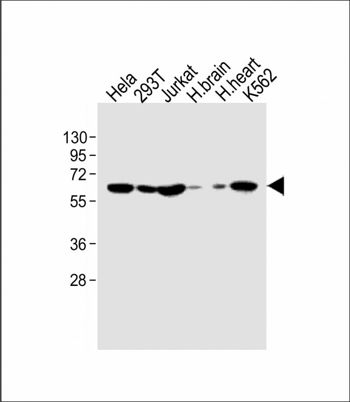
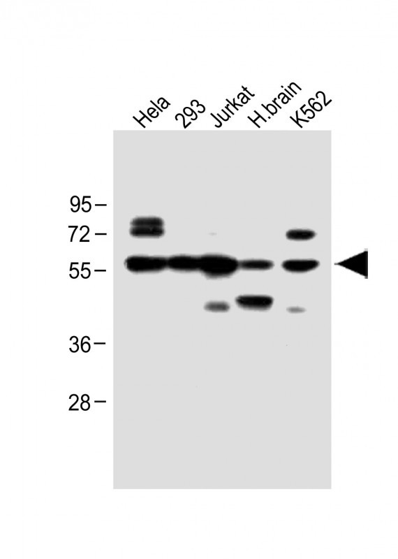
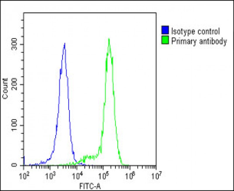
IFNAR1 Antibody (Center)
Affinity Purified Rabbit Polyclonal Antibody (Pab)

- SPECIFICATION
- CITATIONS: 2
- PROTOCOLS
- BACKGROUND

Application
| WB, FC, IHC-P-Leica, E |
|---|---|
| Primary Accession | P17181 |
| Reactivity | Human |
| Host | Rabbit |
| Clonality | Polyclonal |
| Isotype | Rabbit IgG |
| Calculated MW | 63525 Da |
| Antigen Region | 162-188 aa |
| Gene ID | 3454 |
|---|---|
| Other Names | Interferon alpha/beta receptor 1, IFN-R-1, IFN-alpha/beta receptor 1, Cytokine receptor class-II member 1, Cytokine receptor family 2 member 1, CRF2-1, Type I interferon receptor 1, IFNAR1, IFNAR |
| Target/Specificity | This IFNAR1 antibody is generated from rabbits immunized with a KLH conjugated synthetic peptide between 162-188 amino acids of human IFNAR1. |
| Dilution | WB~~1:1000 FC~~1:25 IHC-P-Leica~~1:1000 E~~Use at an assay dependent concentration. |
| Format | Purified polyclonal antibody supplied in PBS with 0.09% (W/V) sodium azide. This antibody is purified through a protein A column, followed by peptide affinity purification. |
| Storage | Maintain refrigerated at 2-8°C for up to 2 weeks. For long term storage store at -20°C in small aliquots to prevent freeze-thaw cycles. |
| Precautions | IFNAR1 Antibody (Center) is for research use only and not for use in diagnostic or therapeutic procedures. |
| Name | IFNAR1 |
|---|---|
| Synonyms | IFNAR |
| Function | Together with IFNAR2, forms the heterodimeric receptor for type I interferons (including interferons alpha, beta, epsilon, omega and kappa) (PubMed:10049744, PubMed:14532120, PubMed:15337770, PubMed:2153461, PubMed:21854986, PubMed:24075985, PubMed:31270247, PubMed:33252644, PubMed:35442418, PubMed:7813427). Type I interferon binding activates the JAK-STAT signaling cascade, resulting in transcriptional activation or repression of interferon-regulated genes that encode the effectors of the interferon response (PubMed:10049744, PubMed:21854986, PubMed:7665574). Mechanistically, type I interferon- binding brings the IFNAR1 and IFNAR2 subunits into close proximity with one another, driving their associated Janus kinases (JAKs) (TYK2 bound to IFNAR1 and JAK1 bound to IFNAR2) to cross-phosphorylate one another (PubMed:21854986, PubMed:32972995, PubMed:7665574, PubMed:7813427). The activated kinases phosphorylate specific tyrosine residues on the intracellular domains of IFNAR1 and IFNAR2, forming docking sites for the STAT transcription factors (PubMed:21854986, PubMed:32972995, PubMed:7526154, PubMed:7665574, PubMed:7813427). STAT proteins are then phosphorylated by the JAKs, promoting their translocation into the nucleus to regulate expression of interferon-regulated genes (PubMed:19561067, PubMed:21854986, PubMed:32972995, PubMed:7665574, PubMed:7813427, PubMed:9121453). Can also act independently of IFNAR2: form an active IFNB1 receptor by itself and activate a signaling cascade that does not involve activation of the JAK-STAT pathway (By similarity). |
| Cellular Location | [Isoform 1]: Cell membrane; Single-pass type I membrane protein. Late endosome. Lysosome. Note=Interferon binding triggers internalization of the receptor from the cell membrane into endosomes and then into lysosomes. |
| Tissue Location | IFN receptors are present in all tissues and even on the surface of most IFN-resistant cells. Isoform 1, isoform 2 and isoform 3 are expressed in the IFN-alpha sensitive myeloma cell line U266B1. Isoform 2 and isoform 3 are expressed in the IFN-alpha resistant myeloma cell line U266R. Isoform 1 is not expressed in IFN- alpha resistant myeloma cell line U266R. |
Research Areas
Citations ( 0 )

Application Protocols
Provided below are standard protocols that you may find useful for product applications.
Background
IFNAR1 is the receptor for interferons alpha and beta. Binding to type I IFNs triggers tyrosine phosphorylation of a number of proteins including JAKs, TYK2, STAT proteins and IFNR alpha-and beta-subunits themselves.
Abcepta welcomes feedback from its customers.
If you have used an Abcepta product and would like to share how it has performed, please click on the "Submit Review" button and provide the requested information. Our staff will examine and post your review and contact you if needed.
If you have any additional inquiries please email technical services at tech@abcepta.com.
$ 150.00
$ 385.00
Cat# AP8550c
Ordering Information
United States
AlbaniaAustraliaAustriaBelgiumBosnia & HerzegovinaBrazilBulgariaCanadaCentral AmericaChinaCroatiaCyprusCzech RepublicDenmarkEstoniaFinlandFranceGermanyGreeceHong KongHungaryIcelandIndiaIndonesiaIrelandIsraelItalyJapanLatviaLithuaniaLuxembourgMacedoniaMalaysiaMaltaMexicoNetherlandsNew ZealandNorwayPakistanPolandPortugalRomaniaSerbiaSingaporeSlovakiaSloveniaSouth AfricaSouth KoreaSpainSwedenSwitzerlandTaiwanTurkeyUnited KingdomUnited StatesVietnamWorldwideOthers
USA Headquarters
(888) 735-7227 / (858) 622-0099 or (858) 875-1900
Other Products
Shipping Information
Domestic orders (in stock items)
Shipped out the same day. Orders placed after 1 PM (PST) will ship out the next business day.
International orders
Contact your local distributors
