


 Foundational characteristics of cancer include proliferation, angiogenesis, migration, evasion of apoptosis, and cellular immortality. Find key markers for these cellular processes and antibodies to detect them.
 The SUMOplot™ Analysis Program predicts and scores sumoylation sites in your protein. SUMOylation is a post-translational modification involved in various cellular processes, such as nuclear-cytosolic transport, transcriptional regulation, apoptosis, protein stability, response to stress, and progression through the cell cycle.
 The Autophagy Receptor Motif Plotter predicts and scores autophagy receptor binding sites in your protein. Identifying proteins connected to this pathway is critical to understanding the role of autophagy in physiological as well as pathological processes such as development, differentiation, neurodegenerative diseases, stress, infection, and cancer.
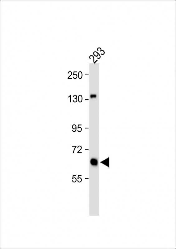
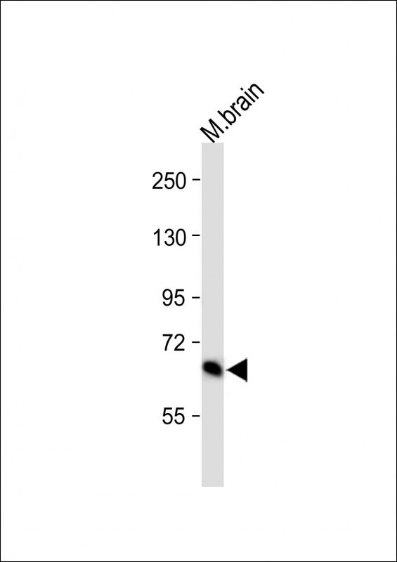
GLS Antibody (C-term)
Affinity Purified Rabbit Polyclonal Antibody (Pab)
- SPECIFICATION
- CITATIONS: 7
- PROTOCOLS
- BACKGROUND

Application
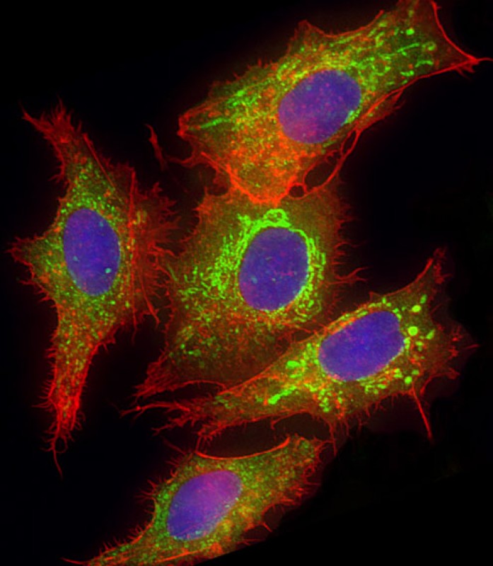
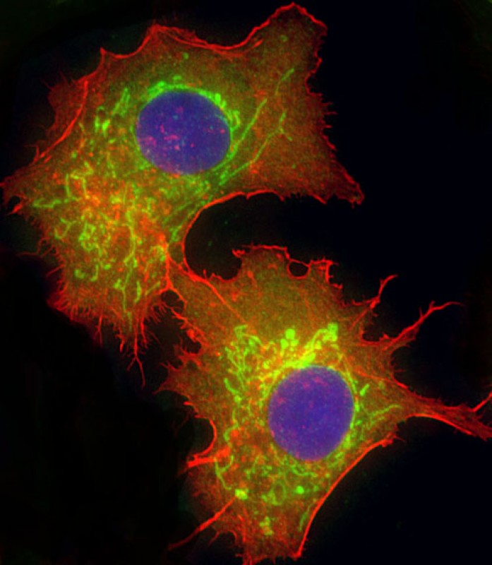
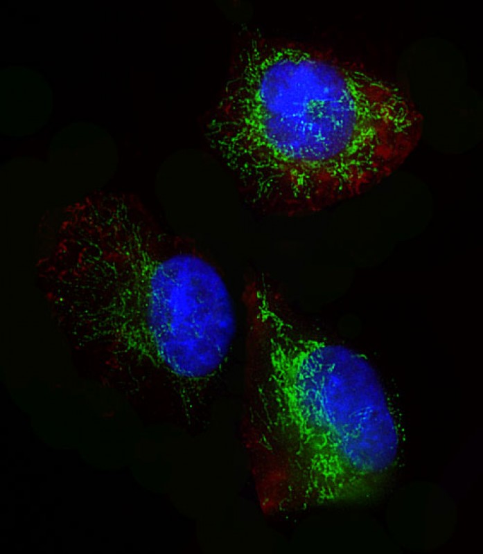
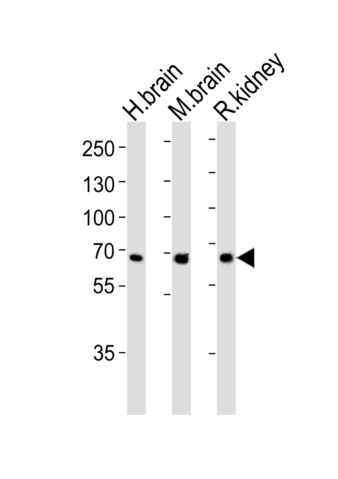
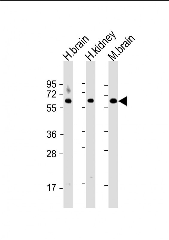
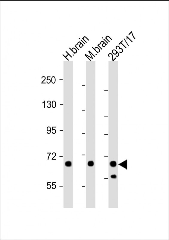
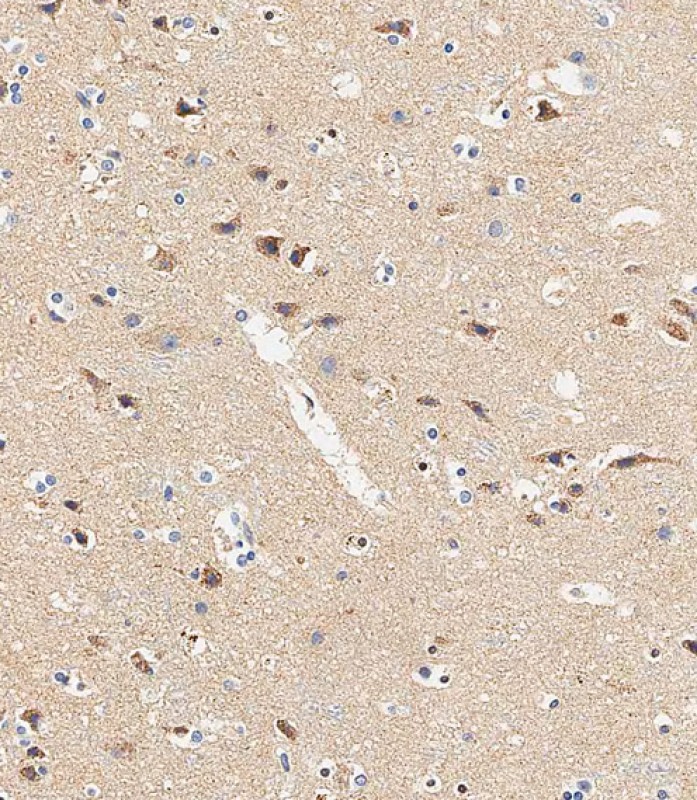
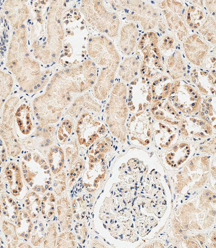
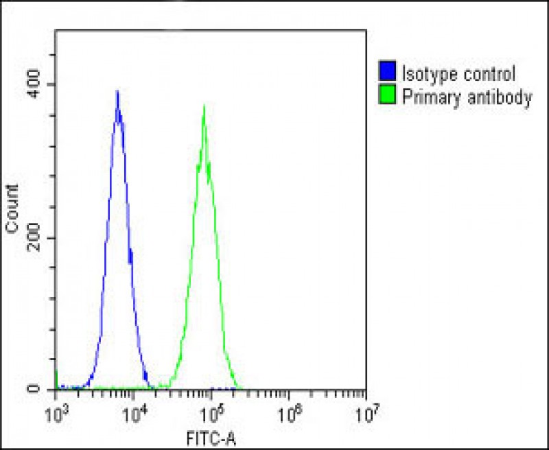
| WB, IHC-P-Leica, IF, FC, E |
|---|---|
| Primary Accession | O94925 |
| Other Accession | P13264, D3Z7P3 |
| Reactivity | Human, Mouse, Rat |
| Predicted | Rat |
| Host | Rabbit |
| Clonality | Polyclonal |
| Isotype | Rabbit IgG |
| Calculated MW | 73461 Da |
| Antigen Region | 516-545 aa |
| Gene ID | 2744 |
|---|---|
| Other Names | Glutaminase kidney isoform, mitochondrial, GLS, K-glutaminase, L-glutamine amidohydrolase, GLS, GLS1, KIAA0838 |
| Target/Specificity | This GLS antibody is generated from rabbits immunized with a KLH conjugated synthetic peptide between 516-545 amino acids from the C-terminal region of human GLS. |
| Dilution | WB~~1:2000 IF~~1:25 IHC-P-Leica~~1:500 FC~~1:25 |
| Format | Purified polyclonal antibody supplied in PBS with 0.09% (W/V) sodium azide. This antibody is purified through a protein A column, followed by peptide affinity purification. |
| Storage | Maintain refrigerated at 2-8°C for up to 2 weeks. For long term storage store at -20°C in small aliquots to prevent freeze-thaw cycles. |
| Precautions | GLS Antibody (C-term) is for research use only and not for use in diagnostic or therapeutic procedures. |
| Name | GLS |
|---|---|
| Synonyms | GLS1, KIAA0838 |
| Function | Catalyzes the first reaction in the primary pathway for the renal catabolism of glutamine. Plays a role in maintaining acid-base homeostasis. Regulates the levels of the neurotransmitter glutamate, the main excitatory neurotransmitter in the brain (PubMed:30575854, PubMed:30239721, PubMed:30970188). |
| Cellular Location | [Isoform 1]: Mitochondrion {ECO:0000250|UniProtKB:P13264}. Cytoplasm, cytosol. Note=The 74-kDa cytosolic precursor is translocated into the mitochondria and processed via a 72-kDa intermediate to yield the mature 68- and 65-kDa subunits {ECO:0000250|UniProtKB:P13264} [Glutaminase kidney isoform, mitochondrial 68 kDa chain]: Mitochondrion matrix {ECO:0000250|UniProtKB:P13264} Note=Produced by the proteolytic processing of the 74-kDa cytosolic precursor. {ECO:0000250|UniProtKB:P13264} |
| Tissue Location | Isoform 1 and isoform 3 are detected in brain cortex. Isoform 3 is highly expressed in astrocytoma, ganglioglioma and ependymoma. Isoform 1 is highly expressed in brain and kidney, but not detected in liver. Isoform 3 is highly expressed in heart and pancreas, detected at lower levels in placenta, lung, pancreas and kidney, but is not detected in liver. Isoform 2 is expressed in cardiac and skeletal muscle. |

Provided below are standard protocols that you may find useful for product applications.
Background
Sahai (1983) demonstrated phosphate-activated glutaminase (EC 3.5.1.2) in human platelets. It is the major enzyme yielding glutamate from glutamine. Significance of the enzyme derives from its possible implication in behavior disturbances in which glutamate acts as a neurotransmitter(Prusiner, 1981). High heritability of platelet glutaminase was indicated by studies of Sahai and Vogel (1983) [PubMed 6682827] who found an intraclass correlation coefficient of 0.96 for monozygotic twins and 0.53 for dizygotic twins.
References
Swierczynski,J., et.al., Biochim. Biophys. Acta 1157 (1), 55-62 (1993)
If you have used an Abcepta product and would like to share how it has performed, please click on the "Submit Review" button and provide the requested information. Our staff will examine and post your review and contact you if needed.
If you have any additional inquiries please email technical services at tech@abcepta.com.
Ordering Information
Other Products
Shipping Information
