


 Foundational characteristics of cancer include proliferation, angiogenesis, migration, evasion of apoptosis, and cellular immortality. Find key markers for these cellular processes and antibodies to detect them.
 The SUMOplot™ Analysis Program predicts and scores sumoylation sites in your protein. SUMOylation is a post-translational modification involved in various cellular processes, such as nuclear-cytosolic transport, transcriptional regulation, apoptosis, protein stability, response to stress, and progression through the cell cycle.
 The Autophagy Receptor Motif Plotter predicts and scores autophagy receptor binding sites in your protein. Identifying proteins connected to this pathway is critical to understanding the role of autophagy in physiological as well as pathological processes such as development, differentiation, neurodegenerative diseases, stress, infection, and cancer.
p33 ING1 Antibody
- SPECIFICATION
- CITATIONS
- PROTOCOLS
- BACKGROUND

Application
| WB, ICC |
|---|---|
| Primary Accession | Q9QXV3 |
| Other Accession | NP_036049.2 |
| Host | Rabbit |
| Reactivity | Human, Mouse |
| Clonality | Polyclonal |
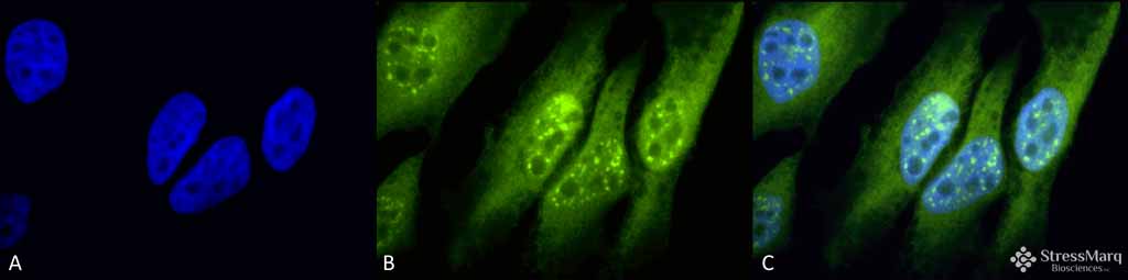
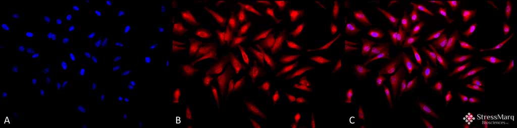
| Description | Rabbit Anti-Human p33 ING1 Polyclonal |
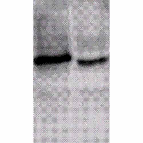
| Target/Specificity | Detects ~33kDa. |
| Other Names | Growth inhibitor ING1 Antibody, ING1 Antibody, inhibitor of growth 1 Antibody, p24ING1c Antibody, p33 Antibody, p33ING Antibody, p33ING1b Antibody, p33ING1c Antibody, p47 Antibody, p47ING1a Antibody, tumor suppressor ING1 Antibody |
| Immunogen | Mouse p33 ING1 N-terminal peptide –KLH conjugates |
| Purification | Protein A Purified |
| Storage | -20ºC |
| Storage Buffer | TBS, 50% glycerol, 0.09% sodium azide |
| Shipping Temperature | Blue Ice or 4ºC |
| Certificate of Analysis | 0.25 mg/ml was sufficient for detection of SPC-145 in lysates prepared from human melanoma cell lines by western blot analysis. |
| Cellular Localization | Nucleus |

Thousands of laboratories across the world have published research that depended on the performance of antibodies from Abcepta to advance their research. Check out links to articles that cite our products in major peer-reviewed journals, organized by research category.
info@abcepta.com, and receive a free "I Love Antibodies" mug.

Provided below are standard protocols that you may find useful for product applications.
Background
The p33ING1 protein encodes a 33-kD, 294-amino acid protein that displays the characteristics of a tumor suppressor gene. The p33ING1 protein is a regulator of cell cycle, senescence, and apoptosis. It can mediate growth arrest, anchor dependent growth and influence chemo sensitivity (1-4). It also has a proposed role as a growth regulator, which is consistent with its location in the nucleus (5). It has been demonstrated that the ING1 gene has three exons and that four mRNA variants are transcribed from three different promoter regions (p33 ING1, p24 ING1, p27 ING1, p47 IG1) (6, 7). ING1 can possibly influence the above biological functions depending on which variant is expressed (1-4).
References
1. Cheung, K.J., Jr. and Li, G. (2002) Int. J. Oncol. 21(6): 1361-1365.
2. Cheung, K.J., Jr. and Li, G. (2002) Exp. Cell Res. 279(2): 291-28.
3. Cheung, K.J., Jr. and Li, G. (2002) Int. J. Oncol. 20(6):1319-1322.
4. Cheung, K.J., Jr. and Li, G. (2001) Exp. Cell Res. 268(1): 1-6.
5. Garkavtsev, I. et al. (1996). Nat Genet. 14(4): 415-420.
6. Gunduz, M. et al. (2000). Cancer Res. 60(12): 3143-6.
7. Jager, D. et al. (1999). Cancer Res. 59: 6197-6204.
If you have used an Abcepta product and would like to share how it has performed, please click on the "Submit Review" button and provide the requested information. Our staff will examine and post your review and contact you if needed.
If you have any additional inquiries please email technical services at tech@abcepta.com.
Ordering Information
Other Products
Shipping Information
