


 Foundational characteristics of cancer include proliferation, angiogenesis, migration, evasion of apoptosis, and cellular immortality. Find key markers for these cellular processes and antibodies to detect them.
 The SUMOplot™ Analysis Program predicts and scores sumoylation sites in your protein. SUMOylation is a post-translational modification involved in various cellular processes, such as nuclear-cytosolic transport, transcriptional regulation, apoptosis, protein stability, response to stress, and progression through the cell cycle.
 The Autophagy Receptor Motif Plotter predicts and scores autophagy receptor binding sites in your protein. Identifying proteins connected to this pathway is critical to understanding the role of autophagy in physiological as well as pathological processes such as development, differentiation, neurodegenerative diseases, stress, infection, and cancer.
Calnexin-CT Antibody

- SPECIFICATION
- CITATIONS
- PROTOCOLS
- BACKGROUND

Application
| WB, IHC, IP, FC, ICC |
|---|---|
| Primary Accession | P24643 |
| Other Accession | NP_001003232.1 |
| Host | Rabbit |
| Reactivity | Human, Mouse, Rat, Rabbit, Hamster, Monkey, Pig, Chicken, Quail, Bovine, Xenopus, Dog, Sheep, Guinea Pig, Drosophila |
| Clonality | Polyclonal |
| Description | Rabbit Anti-Dog Calnexin-CT Polyclonal |
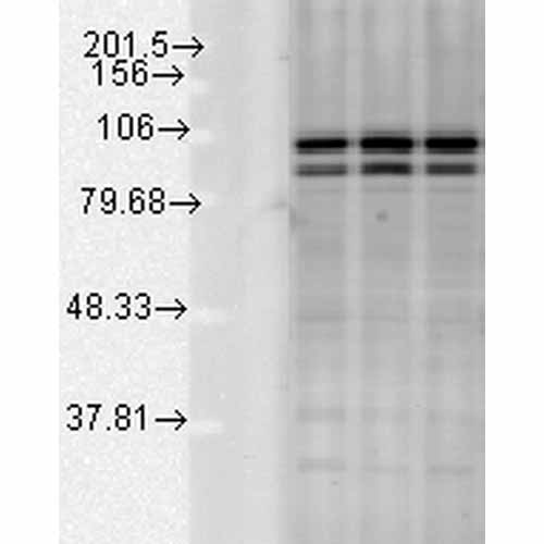
| Target/Specificity | Detects the C-terminal domain of Calnexin ~90kDa. Weak detection in Chicken, Drosophila, and Xenopus tissues. |
| Other Names | Calnexin antibody, CALX_HUMAN antibody, CANX antibody, CNX antibody, FLJ26570 antibody, Histocompatibility complex class I antigen binding protein p88 antibody, IP90 antibody, Major histocompatibility complex class I antigen-binding protein p88 antibody, P90 antibody |
| Immunogen | Dog Calnexin C-terminal synthetic peptide conjugated to KLH. Identical to human, mouse and rat calnexin sequences over these residues. |
| Purification | Protein A Purified |
| Storage | -20ºC |
| Storage Buffer | PBS pH7.2, 50% glycerol, 0.09% sodium azide |
| Shipping Temperature | Blue Ice or 4ºC |
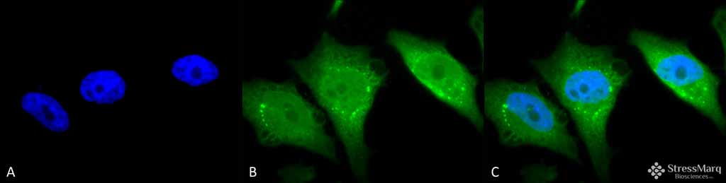
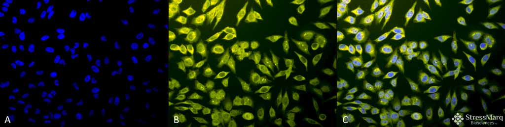
| Certificate of Analysis | A 1:2000 dilution of SPC-182 was sufficient for detection of Calnexin in 10 µg of HeLa cell lysate by ECL immunoblot analysis. |
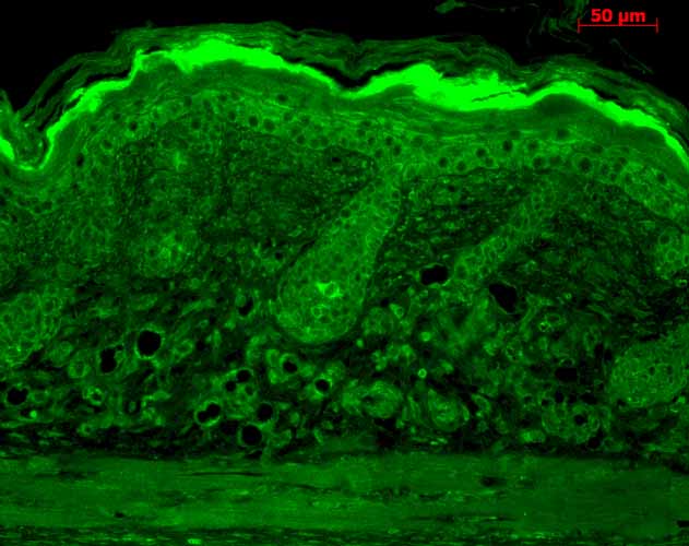
| Cellular Localization | Endoplasmic Reticulum | Endoplasmic Reticulum Membrane | Melanosome |

Thousands of laboratories across the world have published research that depended on the performance of antibodies from Abcepta to advance their research. Check out links to articles that cite our products in major peer-reviewed journals, organized by research category.
info@abcepta.com, and receive a free "I Love Antibodies" mug.

Provided below are standard protocols that you may find useful for product applications.
Background
Calnexin, an abundant ~90kDa integral protein of the endoplasmic reticulum, is also referred to as IP90, p88 and p90 (1). It consists of a large 50kDa N-terminal calcium-binding luminal domain, a single transmembrane helix and a short acidic cytoplasmic tail (2, 3). Unlike its ER counterparts which have a KDEL sequence on their C-terminus to ensure ER retention (4), calnexin has positively charged cytosolic residues that do the same thing (3). Most ER proteins act as molecular chaperones and participate in the proper folding of polypeptides and their assembly into multi-subunit proteins. Calnexin together with calreticulin, plays a key role in glycoprotein folding and its control within the ER, by interacting with folding intermediates via their mono-glycosylated glycans (5, 6). Calnexin has also been shown to associate with the major histocompatibility complex class I heavy chains, partial complexes of the T cell receptor and B cell membrane immunoglobulin (7).
References
1. Rajagopalan S., Xu Y., and Brenner M.B. (1994) Science. 263(5145): 387-90.
2. Tjoelker L.W., et al. (1994) Biochemistry. 33: 3229.
3. Schrag J. et al. (2001) Molecular Cell. 8(3): 633-644.
4. Janiszewski M. (2005) J. Biol Chem. 280(49): 40813-40819.
5. Elagoz A., Callejo M., Armstrong J., and Rokeach L. A. (1999) J. Cell Sci. 112: 4449-4460.
6. Otteken A. and Moss B. (1996) J Bio Chem. 271(1): 97-103.
7. Galvin K. et al. (1992) Proc Natl Acad Sci USA. 89(18): 8452-6.
If you have used an Abcepta product and would like to share how it has performed, please click on the "Submit Review" button and provide the requested information. Our staff will examine and post your review and contact you if needed.
If you have any additional inquiries please email technical services at tech@abcepta.com.
Ordering Information
Other Products
Shipping Information
