


 Foundational characteristics of cancer include proliferation, angiogenesis, migration, evasion of apoptosis, and cellular immortality. Find key markers for these cellular processes and antibodies to detect them.
 The SUMOplot™ Analysis Program predicts and scores sumoylation sites in your protein. SUMOylation is a post-translational modification involved in various cellular processes, such as nuclear-cytosolic transport, transcriptional regulation, apoptosis, protein stability, response to stress, and progression through the cell cycle.
 The Autophagy Receptor Motif Plotter predicts and scores autophagy receptor binding sites in your protein. Identifying proteins connected to this pathway is critical to understanding the role of autophagy in physiological as well as pathological processes such as development, differentiation, neurodegenerative diseases, stress, infection, and cancer.
HSP70 (P. falciparum) Antibody
- SPECIFICATION
- CITATIONS
- PROTOCOLS
- BACKGROUND

Application
| WB, ICC |
|---|---|
| Primary Accession | P11144 |
| Other Accession | M19753 |
| Host | Rabbit |
| Reactivity | P. falciparum |
| Clonality | Polyclonal |
| Description | Rabbit Anti-P. falciparum HSP70 (P. falciparum) Polyclonal |
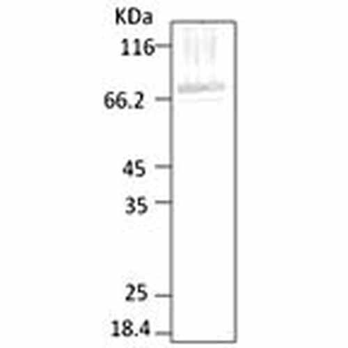
| Target/Specificity | Detects ~ 70kDa. Specific to P. falciparum and does not cross-react to any protein from Human erythrocytes. |
| Other Names | HSP70_PLAFA Antibody, Cytoplasmic antigen 74.3 kDa protein Antibody |
| Immunogen | His-tagged and purified PfHSP70, C-terminus (AA 365-681) |
| Purification | Protein A Purified |
| Storage | -20ºC |
| Storage Buffer | PBS pH7.4, 50% glycerol, 0.09% sodium azide |
| Shipping Temperature | Blue Ice or 4ºC |
| Certificate of Analysis | 0.15 µg/ml of SPC-186 was sufficient for detection of PfHSP70 in 20 µg of P. falciparum lysate by colorimetric immunoblot analysis using Goat anti-rabbit IgG:HRP as the secondary antibody. |
| Cellular Localization | Cytoplasm |

Thousands of laboratories across the world have published research that depended on the performance of antibodies from Abcepta to advance their research. Check out links to articles that cite our products in major peer-reviewed journals, organized by research category.
info@abcepta.com, and receive a free "I Love Antibodies" mug.

Provided below are standard protocols that you may find useful for product applications.
Background
HSP70 genes encode abundant heat-inducible 70-kDa HSPs (HSP70s). In most eukaryotes HSP70 genes exist as part of a multigene family. They are found in most cellular compartments of eukaryotes including nuclei, mitochondria, chloroplasts, the endoplasmic reticulum and the cytosol, as well as in bacteria. The genes show a high degree of conservation, having at least 5O% identity (1). The N-terminal two thirds of HSP70s are more conserved than the C-terminal third. HSP70 binds ATP with high affinity and possesses a weak ATPase activity which can be stimulated by binding to unfolded proteins and synthetic peptides (2). When HSC70 (constitutively expressed) present in mammalian cells was truncated, ATP binding activity was found to reside in an N-terminal fragment of 44 kDa which lacked peptide binding capacity. Polypeptide binding ability therefore resided within the C-terminal half (3). The structure of this ATP binding domain displays multiple features of nucleotide binding proteins (4). All HSP70s, regardless of location, bind proteins, particularly unfolded ones. The molecular chaperones of the HSP70 family recognize and bind to nascent polypeptide chains as well as partially folded intermediates of proteins preventing their aggregation and misfolding. The binding of ATP triggers a critical conformational change leading to the release of the bound substrate protein (5). The universal ability of HSP70s to undergo cycles of binding to and release from hydrophobic stretches of partially unfolded proteins determines their role in a great variety of vital intracellular functions such as protein synthesis, protein folding and oligomerization and protein transport. PfHSP70-I (PF08_0054) is the major cytosolic HSP70 in Plasmodium falciparum. It is abundantly expressed in the blood stages of the parasite and is thought to constitute 1-2% of total parasite protein. It is induced upon heat shock. It is present in the parasite in different complexes with PfHSP90 and some PfHSP40 (6, 7). Looking for more information on HSP70? Visit our new HSP70 Scientific Resource Guide at http://www.HSP70.com.
References
1. Boorstein W. R., Ziegelhoffer T. & Craig E. A. (1993) J. Mol. Evol.38 (1) 1-17.
2. Rothman J. (1989) Cell 59: 591 -601.
3. DeLuca-Flaherty, et al. (1990) Cell. 62: 875-887.
4. Bork P., Sander C. & Valencia A. (1992) Proc. Natl Acad. Sci. USA. 89: 7290-7294.
5. Fink A.L. (1999) Physiol. Rev. 79: 425-449.
6. Pesce E.R., et al. (2008) Int J Biochem Cell Biol. 40(12): 2914-26.
7. Pavithra S.R, Banumathy G., Joy O., Singh V., Tatu U. (2004) J Biol Chem. 279(45): 46692-9.
If you have used an Abcepta product and would like to share how it has performed, please click on the "Submit Review" button and provide the requested information. Our staff will examine and post your review and contact you if needed.
If you have any additional inquiries please email technical services at tech@abcepta.com.
Ordering Information
Shipping Information
