


 Foundational characteristics of cancer include proliferation, angiogenesis, migration, evasion of apoptosis, and cellular immortality. Find key markers for these cellular processes and antibodies to detect them.
 The SUMOplot™ Analysis Program predicts and scores sumoylation sites in your protein. SUMOylation is a post-translational modification involved in various cellular processes, such as nuclear-cytosolic transport, transcriptional regulation, apoptosis, protein stability, response to stress, and progression through the cell cycle.
 The Autophagy Receptor Motif Plotter predicts and scores autophagy receptor binding sites in your protein. Identifying proteins connected to this pathway is critical to understanding the role of autophagy in physiological as well as pathological processes such as development, differentiation, neurodegenerative diseases, stress, infection, and cancer.
AHA2 Antibody
- SPECIFICATION
- CITATIONS
- PROTOCOLS
- BACKGROUND

Application
| WB, ICC |
|---|---|
| Primary Accession | Q719I0 |
| Other Accession | NP_689605.1 |
| Host | Rabbit |
| Reactivity | Human, Mouse, Rat |
| Clonality | Polyclonal |
| Description | Rabbit Anti-Human AHA2 Polyclonal |
| Target/Specificity | Cross reacts with Aha1. |
| Other Names | ATPase hydrogen-exporting ATPase 2 Antibody |
| Immunogen | Aha2 |
| Purification | Protein A Purified |
| Storage | -20ºC |
| Storage Buffer | PBS pH7.4, 50% glycerol, 0.09% sodium azide |
| Shipping Temperature | Blue Ice or 4ºC |
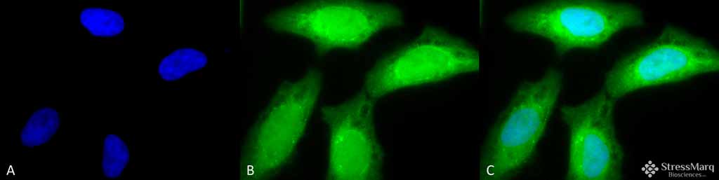
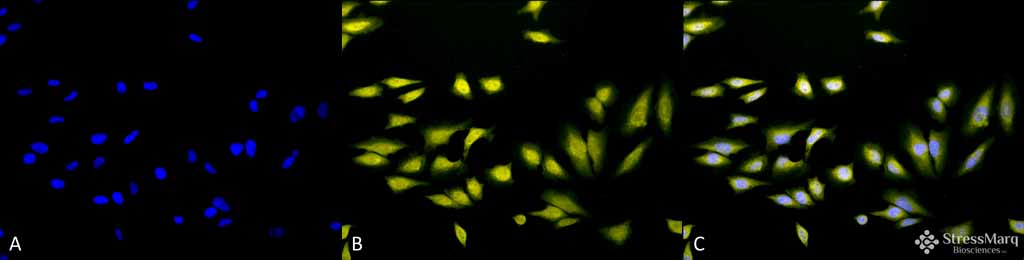
| Certificate of Analysis | 1 µg/ml of SPC-196 was sufficient for detection of Aha2 in 20 µg of rat tissue lysate by colorimetric immunoblot analysis using Goat anti-rat IgG:HRP as the secondary antibody. |

Thousands of laboratories across the world have published research that depended on the performance of antibodies from Abcepta to advance their research. Check out links to articles that cite our products in major peer-reviewed journals, organized by research category.
info@abcepta.com, and receive a free "I Love Antibodies" mug.

Provided below are standard protocols that you may find useful for product applications.
Background
The Arabidopsis protein RIN4 is a well known regulator of plant immunity. The plasma membrane H+-ATPases Aha1 and Aha2 are one class of RIN4-associated proteins (1-5). Aha1 and Aha2 play a crucial role in resisting pathogen invasion- plants use RIN4 to regulate H+-ATPase activity during immune responses, thereby controlling stomatal apertures during pathogen attack (1). Wild type AHA2 has been found to be localized to the plasma membrane, and has also been found in the ER (4).
References
1. Liu J., Elmore J.M., and Coaker G. (2009) Plant Signal Behav. 4(12): 1107-1110.
2. Harper J.F., Surowy T.K. and Sussman M.R. (1989) Proc Natl Acad. Sci USA . 86: 1234-1238.
3. Harper J. F., et al. (1990) J Biol Chem. 265: 13601-13608.
4. Harper J.F., Manney L., and Sussman M.R. (1994) Mol Gen Genet. 243: 572-587.
5. Regenberg B., Villalba J.M., Lanfermeijer F.C., and Palmgren M.G. (1995) Plant Cell. 7: 1655-1666.
If you have used an Abcepta product and would like to share how it has performed, please click on the "Submit Review" button and provide the requested information. Our staff will examine and post your review and contact you if needed.
If you have any additional inquiries please email technical services at tech@abcepta.com.
Ordering Information
Other Products
Shipping Information
