


 Foundational characteristics of cancer include proliferation, angiogenesis, migration, evasion of apoptosis, and cellular immortality. Find key markers for these cellular processes and antibodies to detect them.
 The SUMOplot™ Analysis Program predicts and scores sumoylation sites in your protein. SUMOylation is a post-translational modification involved in various cellular processes, such as nuclear-cytosolic transport, transcriptional regulation, apoptosis, protein stability, response to stress, and progression through the cell cycle.
 The Autophagy Receptor Motif Plotter predicts and scores autophagy receptor binding sites in your protein. Identifying proteins connected to this pathway is critical to understanding the role of autophagy in physiological as well as pathological processes such as development, differentiation, neurodegenerative diseases, stress, infection, and cancer.
Amyloid Fibrils (OC) Antibody
- SPECIFICATION
- CITATIONS
- PROTOCOLS
- BACKGROUND

Application
| WB, IHC, IP, DB, ICC |
|---|---|
| Host | Rabbit |
| Reactivity | Human |
| Clonality | Polyclonal |
| Description | Rabbit Anti-Human Amyloid Fibrils (OC) Polyclonal |
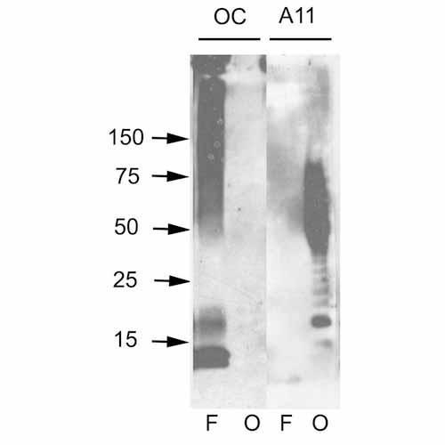
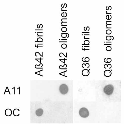
| Target/Specificity | Recognizes generic epitopes common to many amyloid fibrils and fibrillar oligomers, but not prefibrilllar oligomers or natively folded proteins. Expected to detect in Mouse and Rat based on species homology. |
| Other Names | OC Antibody, Fibrils Antibody, Amyloid Oligomer aß Antibody, A11 Antibody, Amyloid beta A4 protein Antibody, ABPP Antibody, APPI Antibody, Alzheimer disease amyloid protein Antibody, Cerebral vascular amyloid peptide Antibody, PreA4 Antibody, Protease nexin-II Antibody, APP Antibody, A4 Antibody, AD Antibody |
| Immunogen | Fibrils prepared from human amyloid beta 42 peptide |
| Purification | Protein A Purified |
| Storage | -20ºC |
| Storage Buffer | PBS, 50% glycerol, 0.09% sodium azide |
| Shipping Temperature | Blue Ice or 4ºC |
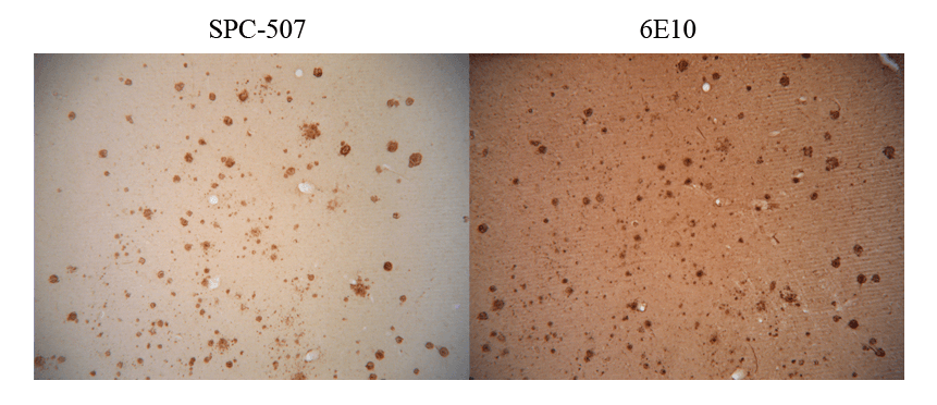
| Certificate of Analysis | A 1:1000 dilution of SPC-507 was sufficient for detection of amyloid fibrils on PVDF membranes using transferred fibrils by colorimetric dot blot analysis using Goat anti-rabbit IgG:HRP as the secondary antibody. |
| Cellular Localization | Membrane |

Thousands of laboratories across the world have published research that depended on the performance of antibodies from Abcepta to advance their research. Check out links to articles that cite our products in major peer-reviewed journals, organized by research category.
info@abcepta.com, and receive a free "I Love Antibodies" mug.

Provided below are standard protocols that you may find useful for product applications.
Background
Amyloid monomeric proteins can sometimes oligomerize into destructive amyloid fibrils. Amyloidogenic conformations of non-disease related proteins can be created by partial protein misfolding or denaturation. Many degenerative diseases are known to be related to the accumulation of misfolded proteins as amyloid fibres (1, 2). These include the amyloid-β peptide plaques and tau neurofibrillary tangles in senile plaques of Alzheimer’s symptomology, the deposition of α-synuclein in the Lewy bodies of Parkinson’s disease, and accumulation of polyglutamine-containing aggregates in Huntington’s disease (2, 3).
References
1. Glabe C.G. (2004) Trends Biochem Sci. 29(10): 542-547.
2. Kayed R., et al. (2004) J Bio. Chem. 279: 46363-46366.
3. Kayed R., et al. (2003) Science. 300(5618): 486-489.
If you have used an Abcepta product and would like to share how it has performed, please click on the "Submit Review" button and provide the requested information. Our staff will examine and post your review and contact you if needed.
If you have any additional inquiries please email technical services at tech@abcepta.com.
Ordering Information
Shipping Information
