


 Foundational characteristics of cancer include proliferation, angiogenesis, migration, evasion of apoptosis, and cellular immortality. Find key markers for these cellular processes and antibodies to detect them.
 The SUMOplot™ Analysis Program predicts and scores sumoylation sites in your protein. SUMOylation is a post-translational modification involved in various cellular processes, such as nuclear-cytosolic transport, transcriptional regulation, apoptosis, protein stability, response to stress, and progression through the cell cycle.
 The Autophagy Receptor Motif Plotter predicts and scores autophagy receptor binding sites in your protein. Identifying proteins connected to this pathway is critical to understanding the role of autophagy in physiological as well as pathological processes such as development, differentiation, neurodegenerative diseases, stress, infection, and cancer.
ULK2 Antibody
- SPECIFICATION
- CITATIONS
- PROTOCOLS
- BACKGROUND

Application
| WB, ICC |
|---|---|
| Primary Accession | Q8IYT8 |
| Other Accession | NP_001136082.1 |
| Host | Rabbit |
| Reactivity | Human, Rat |
| Clonality | Polyclonal |
| Description | Rabbit Anti-Human ULK2 Polyclonal |
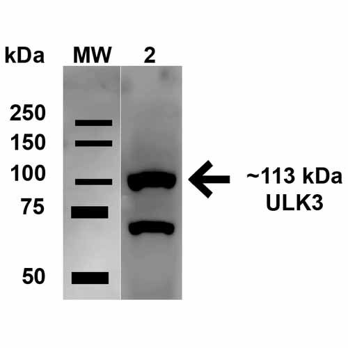
| Target/Specificity | Detects ~100 kDa. |
| Other Names | Serine/threonine-protein kinase ULK2 Antibody, EC:2.7.11.1 Antibody, Unc 51 (C. elegans) like kinase 2 Antibody, ULK2_HUMAN Antibody, KIAA0623 Antibody, ATG1B Antibody, Serine/threonine protein kinase ULK2 Antibody, Unc-51-like kinase 2 Antibody, Unc 51 like autophagy activating kinase 2 Antibody, Unc51.2 Antibody, ULK2 Antibody, Unc 51 like kinase 2 Antibody, |
| Immunogen | Synthetic peptide from the mid-protein of Human ULK2 (aa. 558-569) |
| Purification | Peptide Affinity Purified |
| Storage | -20ºC |
| Storage Buffer | PBS, 50% glycerol, 0.09% sodium azide |
| Shipping Temperature | Blue Ice or 4ºC |
| Certificate of Analysis | A 1:1000 dilution of SPC-641 was sufficient for detection of ULK2 in 15 µg of Human HeLa Cell Lysates by ECL immunoblot analysis using goat anti-rabbit IgG:HRP as the secondary antibody. |
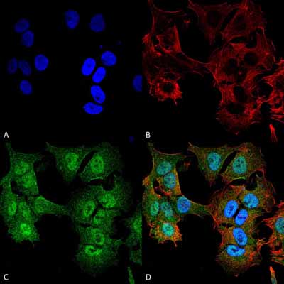
| Cellular Localization | Cytoplasmic Vesicle Membrane | Peripheral Membrane Protein |

Thousands of laboratories across the world have published research that depended on the performance of antibodies from Abcepta to advance their research. Check out links to articles that cite our products in major peer-reviewed journals, organized by research category.
info@abcepta.com, and receive a free "I Love Antibodies" mug.

Provided below are standard protocols that you may find useful for product applications.
Background
UNC-51 like kinase 2 (ULK2) is widely expresed and contains an amino-terminal kinase domain followed by a central proline-serine rich domain and a highly conserved carboxy-terminal domain. It has been linked to axon growth and is essential for autophagy. Structurally, ULK2 is similar to ATG1.
References
1. Okazaki N., et al. (2000) Brain Res Mol Brain Res. 85: 1-12.
2. Young A.R., et al. (2006) J Cell Sci. 119: 3888-900.
3. Kamada Y., et al. (2000) J Cell Biol. 150: 1507-13.
4. Lee S.B, et al. (2007) EMBO Rep. 8: 360-5.
5. Hara T., et al. (2008) J Cell Biol. 181: 497-510.
If you have used an Abcepta product and would like to share how it has performed, please click on the "Submit Review" button and provide the requested information. Our staff will examine and post your review and contact you if needed.
If you have any additional inquiries please email technical services at tech@abcepta.com.
Ordering Information
Other Products
Shipping Information
