


 Foundational characteristics of cancer include proliferation, angiogenesis, migration, evasion of apoptosis, and cellular immortality. Find key markers for these cellular processes and antibodies to detect them.
 The SUMOplot™ Analysis Program predicts and scores sumoylation sites in your protein. SUMOylation is a post-translational modification involved in various cellular processes, such as nuclear-cytosolic transport, transcriptional regulation, apoptosis, protein stability, response to stress, and progression through the cell cycle.
 The Autophagy Receptor Motif Plotter predicts and scores autophagy receptor binding sites in your protein. Identifying proteins connected to this pathway is critical to understanding the role of autophagy in physiological as well as pathological processes such as development, differentiation, neurodegenerative diseases, stress, infection, and cancer.
IRGM Antibody
- SPECIFICATION
- CITATIONS
- PROTOCOLS
- BACKGROUND

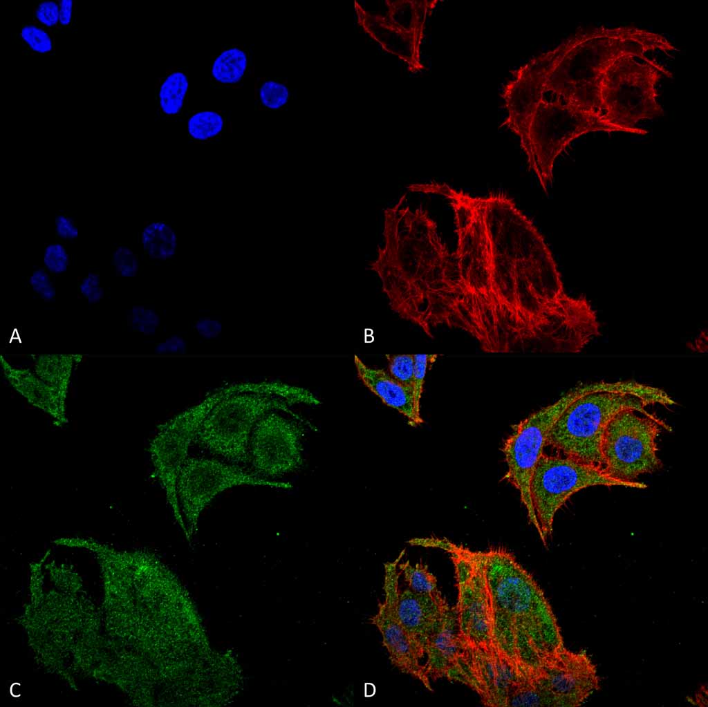
Application
| WB, ICC |
|---|---|
| Primary Accession | A1A4Y4 |
| Other Accession | NP_001139277.1 |
| Host | Rabbit |
| Reactivity | Human |
| Clonality | Polyclonal |
| Description | Rabbit Anti-Human IRGM Polyclonal |
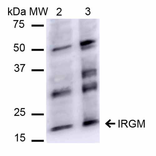
| Target/Specificity | Detects ~20 kDa. |
| Other Names | Immunity related GTPase family M Antibody Iipg3 Antibody Interferon inducible protein 1 Antibody, LRG-47-like protein Antibody, Immunity-related GTPase family M protein 1 Antibody, IFI1 Antibody, Irgm Antibody, IRGM_HUMAN Antibody, LRG47 Antibody, Interferon-inducible protein 1 Antibody, IRGM1 Antibody, LRG 47 Antibody, IRGM Antibody, Iigp3 Antibody, Immunity-related GTPase family M protein Antibody, LRG-47 Antibody, MGC149263 Antibody, Immunity related GTPase family M protein 1 Antibody, LPS-stimulated RAW 264.7 macrophage protein 47 homolog Antibody |
| Immunogen | Synthetic peptide from the N-terminal of Human IRGM |
| Purification | Peptide Affinity Purified |
| Storage | -20ºC |
| Storage Buffer | PBS, 50% glycerol, 0.09% sodium azide |
| Shipping Temperature | Blue Ice or 4ºC |
| Certificate of Analysis | A 1:1000 dilution of SPC-681 was sufficient for detection of IRGM in 15 µg of human HeLa cell lysates by ECL immunoblot analysis using goat anti-rabbit IgG:HRP as the secondary antibody. |
| Cellular Localization | Golgi Apparatus Membrane | Cell Membrane | Cytoplasmic Vesicle | Phagosome Membrane | Cytoplasmic Vesicle | Autophagosome Membrane | Cell Projection | Phagocytic Cup |
Research Areas
Citations (0)

Thousands of laboratories across the world have published research that depended on the performance of antibodies from Abcepta to advance their research. Check out links to articles that cite our products in major peer-reviewed journals, organized by research category.
Submit your citation using an Abcepta antibody to
info@abcepta.com, and receive a free "I Love Antibodies" mug.
info@abcepta.com, and receive a free "I Love Antibodies" mug.

Application Protocols
Provided below are standard protocols that you may find useful for product applications.
Abcepta welcomes feedback from its customers.
If you have used an Abcepta product and would like to share how it has performed, please click on the "Submit Review" button and provide the requested information. Our staff will examine and post your review and contact you if needed.
If you have any additional inquiries please email technical services at tech@abcepta.com.
Cat# ASM10541
Ordering Information
United States
AlbaniaAustraliaAustriaBelgiumBosnia & HerzegovinaBrazilBulgariaCanadaCentral AmericaChinaCroatiaCyprusCzech RepublicDenmarkEstoniaFinlandFranceGermanyGreeceHong KongHungaryIcelandIndiaIndonesiaIrelandIsraelItalyJapanLatviaLithuaniaLuxembourgMacedoniaMalaysiaMaltaMexicoNetherlandsNew ZealandNorwayPakistanPolandPortugalRomaniaSerbiaSingaporeSlovakiaSloveniaSouth AfricaSouth KoreaSpainSwedenSwitzerlandTaiwanTurkeyUnited KingdomUnited StatesVietnamWorldwideOthers
USA Headquarters
(888) 735-7227 / (858) 622-0099 or (858) 875-1900
Other Products
Shipping Information
Domestic orders (in stock items)
Shipped out the same day. Orders placed after 1 PM (PST) will ship out the next business day.
International orders
Contact your local distributors
