


 Foundational characteristics of cancer include proliferation, angiogenesis, migration, evasion of apoptosis, and cellular immortality. Find key markers for these cellular processes and antibodies to detect them.
 The SUMOplot™ Analysis Program predicts and scores sumoylation sites in your protein. SUMOylation is a post-translational modification involved in various cellular processes, such as nuclear-cytosolic transport, transcriptional regulation, apoptosis, protein stability, response to stress, and progression through the cell cycle.
 The Autophagy Receptor Motif Plotter predicts and scores autophagy receptor binding sites in your protein. Identifying proteins connected to this pathway is critical to understanding the role of autophagy in physiological as well as pathological processes such as development, differentiation, neurodegenerative diseases, stress, infection, and cancer.
BNIP3 Antibody

- SPECIFICATION
- CITATIONS
- PROTOCOLS
- BACKGROUND

Application
| WB, ICC |
|---|---|
| Primary Accession | Q12983 |
| Other Accession | NP_004043.3 |
| Host | Rabbit |
| Reactivity | Human |
| Clonality | Polyclonal |
| Description | Rabbit Anti-Human BNIP3 Polyclonal |
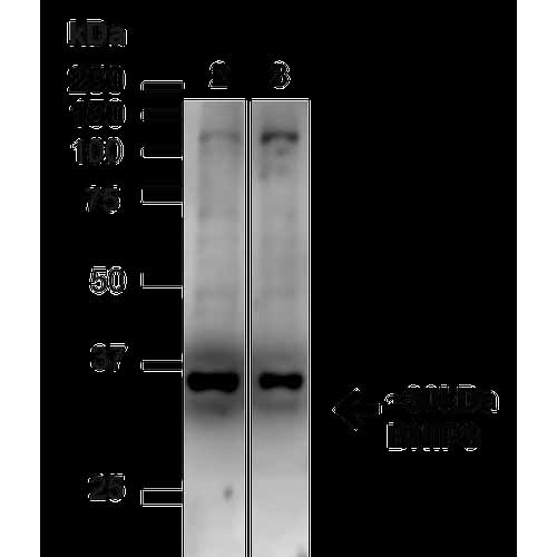
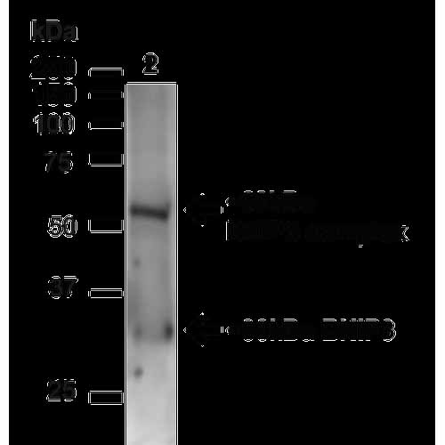
| Target/Specificity | Predicted molecular weight at ~21.5kDa. Observed bands at ~20-30kDa and 50-60kDa due to the homodimer complex of BNIP3. |
| Other Names | NIP3 Antibody, BCL2 Adenovirus E1B 19kDa Interacting Protein 3 Antibody, BNIP3_Human Antibody |
| Immunogen | Synthetic peptide from the C-terminal of human BNIP3 |
| Purification | Peptide Affinity Purified |
| Storage | -20ºC |
| Storage Buffer | PBS, 50% glycerol, 0.09% sodium azide |
| Shipping Temperature | Blue Ice or 4ºC |
| Certificate of Analysis | A 1:1000 dilution of SPC-685 was sufficient for detection of BNIP3 on mouse kidney lysates using Goat anti-rabbit IgG:HRP as the secondary antibody. |
| Cellular Localization | Mitochondrion | Mitochondrion outer membrane |

Thousands of laboratories across the world have published research that depended on the performance of antibodies from Abcepta to advance their research. Check out links to articles that cite our products in major peer-reviewed journals, organized by research category.
info@abcepta.com, and receive a free "I Love Antibodies" mug.

Provided below are standard protocols that you may find useful for product applications.
Background
BNIP3 is an apoptosis-inducing protein which can overcome BCL2 suppression. It may play a role in repartioning calcium between the two major intracellular calcium stores in association with BCL2. It has been shown to interact with CD47, BCL2-like, and BCL-2 (1-3).
References
1. Boyd J.M., et al. (1994) Cell. 79(2): 341-351.
2. Lamy L., et al. (2003) J Biol Chem. 278(26): 23915-23921.
3. Ray R., et al. (2000) J Biol Chem. 275(2): 1439-1448.
If you have used an Abcepta product and would like to share how it has performed, please click on the "Submit Review" button and provide the requested information. Our staff will examine and post your review and contact you if needed.
If you have any additional inquiries please email technical services at tech@abcepta.com.
Ordering Information
Other Products
Shipping Information
