


 Foundational characteristics of cancer include proliferation, angiogenesis, migration, evasion of apoptosis, and cellular immortality. Find key markers for these cellular processes and antibodies to detect them.
 The SUMOplot™ Analysis Program predicts and scores sumoylation sites in your protein. SUMOylation is a post-translational modification involved in various cellular processes, such as nuclear-cytosolic transport, transcriptional regulation, apoptosis, protein stability, response to stress, and progression through the cell cycle.
 The Autophagy Receptor Motif Plotter predicts and scores autophagy receptor binding sites in your protein. Identifying proteins connected to this pathway is critical to understanding the role of autophagy in physiological as well as pathological processes such as development, differentiation, neurodegenerative diseases, stress, infection, and cancer.
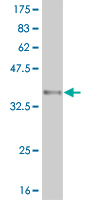
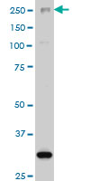
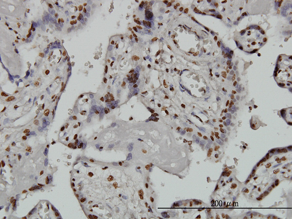
ARID1B Antibody (monoclonal) (M02)
Mouse monoclonal antibody raised against a partial recombinant ARID1B.

- SPECIFICATION
- CITATIONS: 4
- PROTOCOLS
- BACKGROUND

Application
| WB, IHC, IF |
|---|---|
| Primary Accession | Q8NFD5 |
| Other Accession | NM_017519 |
| Reactivity | Human |
| Host | Mouse |
| Clonality | Monoclonal |
| Isotype | IgG2b Kappa |
| Clone Names | 2F2 |
| Calculated MW | 243943 Da |
| Gene ID | 57492 |
|---|---|
| Other Names | AT-rich interactive domain-containing protein 1B, ARID domain-containing protein 1B, BRG1-associated factor 250b, BAF250B, BRG1-binding protein hELD/OSA1, Osa homolog 2, hOsa2, p250R, ARID1B, BAF250B, DAN15, KIAA1235, OSA2 |
| Target/Specificity | ARID1B (NP_059989, 1364 a.a. ~ 1460 a.a) partial recombinant protein with GST tag. MW of the GST tag alone is 26 KDa. |
| Dilution | WB~~1:500~1000 IHC~~1:100~500 IF~~1:50~200 |
| Format | Clear, colorless solution in phosphate buffered saline, pH 7.2 . |
| Storage | Store at -20°C or lower. Aliquot to avoid repeated freezing and thawing. |
| Precautions | ARID1B Antibody (monoclonal) (M02) is for research use only and not for use in diagnostic or therapeutic procedures. |
Citations ( 0 )

Application Protocols
Provided below are standard protocols that you may find useful for product applications.
References
1.Dynamics of expression of ARID1A and ARID1B subunits in mouse embryos and in cells during the cell cycle.Flores-Alcantar A, Gonzalez-Sandoval A, Escalante-Alcalde D, Lomeli H.Cell Tissue Res. 2011 Jun 7. [Epub ahead of print]
Abcepta welcomes feedback from its customers.
If you have used an Abcepta product and would like to share how it has performed, please click on the "Submit Review" button and provide the requested information. Our staff will examine and post your review and contact you if needed.
If you have any additional inquiries please email technical services at tech@abcepta.com.
$ 350.00
Cat# AT1190a
Ordering Information
United States
AlbaniaAustraliaAustriaBelgiumBosnia & HerzegovinaBrazilBulgariaCanadaCentral AmericaChinaCroatiaCyprusCzech RepublicDenmarkEstoniaFinlandFranceGermanyGreeceHong KongHungaryIcelandIndiaIndonesiaIrelandIsraelItalyJapanLatviaLithuaniaLuxembourgMacedoniaMalaysiaMaltaMexicoNetherlandsNew ZealandNorwayPakistanPolandPortugalRomaniaSerbiaSingaporeSlovakiaSloveniaSouth AfricaSouth KoreaSpainSwedenSwitzerlandTaiwanTurkeyUnited KingdomUnited StatesVietnamWorldwideOthers
USA Headquarters
(888) 735-7227 / (858) 622-0099 or (858) 875-1900
Other Products
Shipping Information
Domestic orders (in stock items)
Shipped out the same day. Orders placed after 1 PM (PST) will ship out the next business day.
International orders
Contact your local distributors
