


 Foundational characteristics of cancer include proliferation, angiogenesis, migration, evasion of apoptosis, and cellular immortality. Find key markers for these cellular processes and antibodies to detect them.
 The SUMOplot™ Analysis Program predicts and scores sumoylation sites in your protein. SUMOylation is a post-translational modification involved in various cellular processes, such as nuclear-cytosolic transport, transcriptional regulation, apoptosis, protein stability, response to stress, and progression through the cell cycle.
 The Autophagy Receptor Motif Plotter predicts and scores autophagy receptor binding sites in your protein. Identifying proteins connected to this pathway is critical to understanding the role of autophagy in physiological as well as pathological processes such as development, differentiation, neurodegenerative diseases, stress, infection, and cancer.
TCF4 Antibody (monoclonal) (M01)
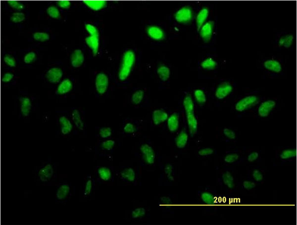
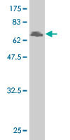
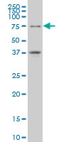
Mouse monoclonal antibody raised against a full length recombinant TCF4.
- SPECIFICATION
- CITATIONS: 1
- PROTOCOLS
- BACKGROUND

Application
| WB, IF |
|---|---|
| Primary Accession | P15884 |
| Other Accession | BC031056.1 |
| Reactivity | Human |
| Host | mouse |
| Clonality | Monoclonal |
| Isotype | IgG1 Kappa |
| Clone Names | 3E11 |
| Calculated MW | 71308 Da |
| Gene ID | 6925 |
|---|---|
| Other Names | Transcription factor 4, TCF-4, Class B basic helix-loop-helix protein 19, bHLHb19, Immunoglobulin transcription factor 2, ITF-2, SL3-3 enhancer factor 2, SEF-2, TCF4, BHLHB19, ITF2, SEF2 |
| Target/Specificity | TCF4 (AAH31056.1, 1 a.a. ~ 365 a.a) full-length recombinant protein with GST tag. MW of the GST tag alone is 26 KDa. |
| Dilution | WB~~1:500~1000 IF~~1:50~200 |
| Format | Clear, colorless solution in phosphate buffered saline, pH 7.2 . |
| Storage | Store at -20°C or lower. Aliquot to avoid repeated freezing and thawing. |
| Precautions | TCF4 Antibody (monoclonal) (M01) is for research use only and not for use in diagnostic or therapeutic procedures. |

Provided below are standard protocols that you may find useful for product applications.
Background
This gene encodes transcription factor 4, a basic helix-turn-helix transcription factor. The encoded protein recognizes an Ephrussi-box ('E-box') binding site ('CANNTG') - a motif first identified in immunoglobulin enhancers. This gene is expressed predominantly in pre-B-cells, although it is found in other tissues as well. Multiple alternatively spliced transcript variants that encode different proteins have been described. [provided by RefSeq]
If you have used an Abcepta product and would like to share how it has performed, please click on the "Submit Review" button and provide the requested information. Our staff will examine and post your review and contact you if needed.
If you have any additional inquiries please email technical services at tech@abcepta.com.
Ordering Information
Other Products
Shipping Information
