


 Foundational characteristics of cancer include proliferation, angiogenesis, migration, evasion of apoptosis, and cellular immortality. Find key markers for these cellular processes and antibodies to detect them.
 The SUMOplot™ Analysis Program predicts and scores sumoylation sites in your protein. SUMOylation is a post-translational modification involved in various cellular processes, such as nuclear-cytosolic transport, transcriptional regulation, apoptosis, protein stability, response to stress, and progression through the cell cycle.
 The Autophagy Receptor Motif Plotter predicts and scores autophagy receptor binding sites in your protein. Identifying proteins connected to this pathway is critical to understanding the role of autophagy in physiological as well as pathological processes such as development, differentiation, neurodegenerative diseases, stress, infection, and cancer.
MC1R Antibody (Center)
Mouse Monoclonal Antibody (Mab)
- SPECIFICATION
- CITATIONS
- PROTOCOLS
- BACKGROUND

Application
| WB, E |
|---|---|
| Primary Accession | Q01726 |
| Other Accession | NP_002377.4 |
| Reactivity | Human |
| Host | Mouse |
| Clonality | Monoclonal |
| Isotype | IgM |
| Clone/Animal Names | 578CT6.2.3 |
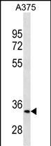
| Calculated MW | 34706 Da |
| Antigen Region | 205-232 aa |
| Gene ID | 4157 |
|---|---|
| Other Names | Melanocyte-stimulating hormone receptor, MSH-R, Melanocortin receptor 1, MC1-R, MC1R, MSHR |
| Target/Specificity | This MC1R antibody is generated from mice immunized with a KLH conjugated synthetic peptide between 205-232 amino acids from the Central region of human MC1R. |
| Dilution | WB~~1:500~1000 E~~Use at an assay dependent concentration. |
| Format | Purified monoclonal antibody supplied in PBS with 0.09% (W/V) sodium azide. This antibody is prepared by Euglobin precipitation followed by dialysis against PBS. |
| Storage | Maintain refrigerated at 2-8°C for up to 2 weeks. For long term storage store at -20°C in small aliquots to prevent freeze-thaw cycles. |
| Precautions | MC1R Antibody (Center) is for research use only and not for use in diagnostic or therapeutic procedures. |
| Name | MC1R |
|---|---|
| Synonyms | MSHR |
| Function | Receptor for MSH (alpha, beta and gamma) and ACTH (PubMed:11442765, PubMed:11707265, PubMed:1325670, PubMed:1516719, PubMed:8463333). The activity of this receptor is mediated by G proteins which activate adenylate cyclase (PubMed:11707265, PubMed:1325670, PubMed:16463023, PubMed:19737927). Mediates melanogenesis, the production of eumelanin (black/brown) and phaeomelanin (red/yellow), via regulation of cAMP signaling in melanocytes (PubMed:31097585). |
| Cellular Location | Cell membrane; Multi-pass membrane protein |
| Tissue Location | Expressed in melanocytes (PubMed:1325670, PubMed:31097585). Expressed in corticoadrenal tissue (PubMed:1325670) |

Thousands of laboratories across the world have published research that depended on the performance of antibodies from Abcepta to advance their research. Check out links to articles that cite our products in major peer-reviewed journals, organized by research category.
info@abcepta.com, and receive a free "I Love Antibodies" mug.

Provided below are standard protocols that you may find useful for product applications.
Background
This intronless gene encodes the receptor protein for melanocyte-stimulating hormone (MSH). The encoded protein, a seven pass transmembrane G protein coupled receptor, controls melanogenesis. Two types of melanin exist: red pheomelanin and black eumelanin. Gene mutations that lead to a loss in function are associated with increased pheomelanin production, which leads to lighter skin and hair color. Eumelanin is photoprotective but pheomelanin may contribute to UV-induced skin damage by generating free radicals upon UV radiation. Binding of MSH to its receptor activates the receptor and stimulates eumelanin synthesis. This receptor is a major determining factor in sun sensitivity and is a genetic risk factor for melanoma and non-melanoma skin cancer. Over 30 variant alleles have been identified which correlate with skin and hair color, providing evidence that this gene is an important component in determining normal human pigment variation. [provided by RefSeq].
References
Demenais, F., et al. J. Natl. Cancer Inst. 102(20):1568-1583(2010)
Strange, R.C., et al. Mult. Scler. 16(9):1109-1116(2010)
Ibarrola-Villava, M., et al. Exp. Dermatol. 19(9):836-844(2010)
Smith, G., et al. Pharmacogenet. Genomics (2010) In press :
Kricker, A., et al. Cancer Causes Control (2010) In press :
If you have used an Abcepta product and would like to share how it has performed, please click on the "Submit Review" button and provide the requested information. Our staff will examine and post your review and contact you if needed.
If you have any additional inquiries please email technical services at tech@abcepta.com.
Ordering Information
Other Products
Shipping Information
