


 Foundational characteristics of cancer include proliferation, angiogenesis, migration, evasion of apoptosis, and cellular immortality. Find key markers for these cellular processes and antibodies to detect them.
 The SUMOplot™ Analysis Program predicts and scores sumoylation sites in your protein. SUMOylation is a post-translational modification involved in various cellular processes, such as nuclear-cytosolic transport, transcriptional regulation, apoptosis, protein stability, response to stress, and progression through the cell cycle.
 The Autophagy Receptor Motif Plotter predicts and scores autophagy receptor binding sites in your protein. Identifying proteins connected to this pathway is critical to understanding the role of autophagy in physiological as well as pathological processes such as development, differentiation, neurodegenerative diseases, stress, infection, and cancer.
SLC43A2 Antibody (Center)
Affinity Purified Rabbit Polyclonal Antibody (Pab)
- SPECIFICATION
- CITATIONS: 1
- PROTOCOLS
- BACKGROUND

Application
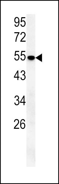
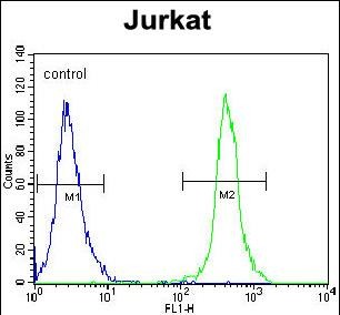
| WB, FC, E |
|---|---|
| Primary Accession | Q8N370 |
| Other Accession | Q8CGA3, Q0VCM6, NP_689559.1 |
| Reactivity | Human, Mouse |
| Predicted | Bovine |
| Host | Rabbit |
| Clonality | Polyclonal |
| Isotype | Rabbit IgG |
| Calculated MW | 62747 Da |
| Antigen Region | 236-265 aa |
| Gene ID | 124935 |
|---|---|
| Other Names | Large neutral amino acids transporter small subunit 4, L-type amino acid transporter 4, Solute carrier family 43 member 2, SLC43A2, LAT4 |
| Target/Specificity | This SLC43A2 antibody is generated from rabbits immunized with a KLH conjugated synthetic peptide between 236-265 amino acids from the Central region of human SLC43A2. |
| Dilution | WB~~1:1000 FC~~1:10~50 E~~Use at an assay dependent concentration. |
| Format | Purified polyclonal antibody supplied in PBS with 0.09% (W/V) sodium azide. This antibody is purified through a protein A column, followed by peptide affinity purification. |
| Storage | Maintain refrigerated at 2-8°C for up to 2 weeks. For long term storage store at -20°C in small aliquots to prevent freeze-thaw cycles. |
| Precautions | SLC43A2 Antibody (Center) is for research use only and not for use in diagnostic or therapeutic procedures. |
| Name | SLC43A2 (HGNC:23087) |
|---|---|
| Synonyms | LAT4 |
| Function | Uniporter that mediates the transport of the stereospecific L-phenylalanine, L-methionine and L-branched-chain amino acids, between the extracellular space and the cytoplasm and may control the transepithelial (re)absorption of neutral amino acid in kidney and small intestine (PubMed:15659399, PubMed:30379325). The transport activity is mediated through facilitated diffusion and is sodium ions-, chloride ions- and pH-independent (PubMed:15659399). |
| Cellular Location | Cell membrane; Multi-pass membrane protein. Basolateral cell membrane {ECO:0000250|UniProtKB:Q8CGA3}. Note=Located at the basolateral membrane in the small intestine enterocytes, kidney proximal tubule, thick ascending limb and, to a minor extent, of distal convoluted tubule epithelial cells. {ECO:0000250|UniProtKB:Q8CGA3} |
| Tissue Location | Detected in several tissues with higher expression in placenta, kidney and peripheral blood leukocytes (PubMed:15659399) In the kidney, is detected in epithelial cells of the distal tubule and collecting duct (PubMed:15659399). In the intestine, is expressed mainly in crypt cells of the intestinal microvilli and epithelial cells in the base of the villus (PubMed:15659399) |
Research Areas
Citations ( 0 )

Application Protocols
Provided below are standard protocols that you may find useful for product applications.
Abcepta welcomes feedback from its customers.
If you have used an Abcepta product and would like to share how it has performed, please click on the "Submit Review" button and provide the requested information. Our staff will examine and post your review and contact you if needed.
If you have any additional inquiries please email technical services at tech@abcepta.com.
$ 150.00
$ 385.00
Cat# AP10062c
Ordering Information
United States
AlbaniaAustraliaAustriaBelgiumBosnia & HerzegovinaBrazilBulgariaCanadaCentral AmericaChinaCroatiaCyprusCzech RepublicDenmarkEstoniaFinlandFranceGermanyGreeceHong KongHungaryIcelandIndiaIndonesiaIrelandIsraelItalyJapanLatviaLithuaniaLuxembourgMacedoniaMalaysiaMaltaMexicoNetherlandsNew ZealandNorwayPakistanPolandPortugalRomaniaSerbiaSingaporeSlovakiaSloveniaSouth AfricaSouth KoreaSpainSwedenSwitzerlandTaiwanTurkeyUnited KingdomUnited StatesVietnamWorldwideOthers
USA Headquarters
(888) 735-7227 / (858) 622-0099 or (858) 875-1900
Other Products
Shipping Information
Domestic orders (in stock items)
Shipped out the same day. Orders placed after 1 PM (PST) will ship out the next business day.
International orders
Contact your local distributors
