


 Foundational characteristics of cancer include proliferation, angiogenesis, migration, evasion of apoptosis, and cellular immortality. Find key markers for these cellular processes and antibodies to detect them.
 The SUMOplot™ Analysis Program predicts and scores sumoylation sites in your protein. SUMOylation is a post-translational modification involved in various cellular processes, such as nuclear-cytosolic transport, transcriptional regulation, apoptosis, protein stability, response to stress, and progression through the cell cycle.
 The Autophagy Receptor Motif Plotter predicts and scores autophagy receptor binding sites in your protein. Identifying proteins connected to this pathway is critical to understanding the role of autophagy in physiological as well as pathological processes such as development, differentiation, neurodegenerative diseases, stress, infection, and cancer.
Erk1/2 Antibody
- SPECIFICATION
- CITATIONS
- PROTOCOLS
- BACKGROUND

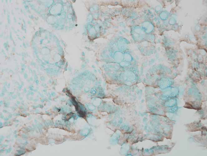
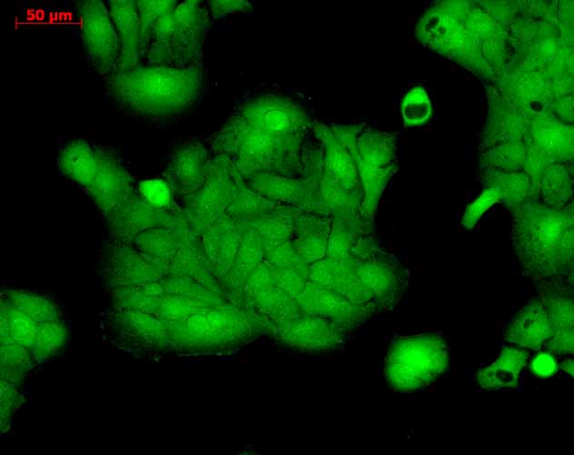
Application
| WB, IHC, FC, ICC |
|---|---|
| Primary Accession | P21708 |
| Other Accession | NP_059043.1 |
| Host | Rabbit |
| Reactivity | Human, Mouse, Rat, Chicken, Bovine, Xenopus, Sheep, Drosophila |
| Clonality | Polyclonal |
| Description | Rabbit Anti-Rat Erk1/2 Polyclonal |
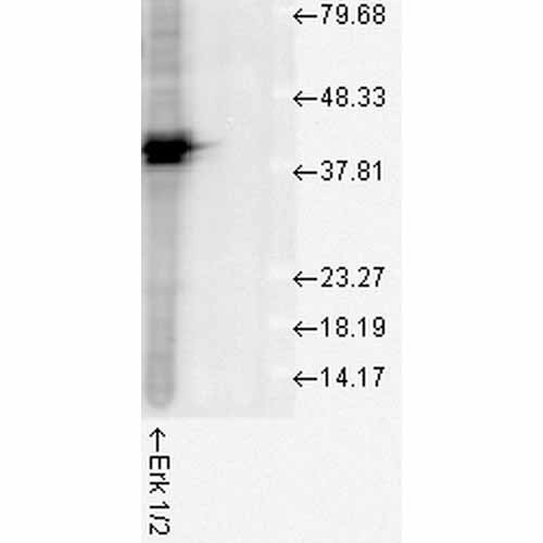
| Target/Specificity | Detects ~44kda (ERK1) and ~42kDa (ERK2). |
| Other Names | ERK1 Antibody, ERK2 Antibody, ERT1 Antibody, ERT2 Antibody, MAP kinase1 Antibody, MAP kinase2 Antibody, MAPK1 Antibody, MAPK2 Antibody, MAPK3 Antibody, p38 Antibody, p40 Antibody, p41 Antibody, p41mapk Antibody, p42 MAPK Antibody, p44 ERK1 Antibody, p44 MAPK Antibody, PRKM1 PRKM2 Antibody, PRKM3 Antibody |
| Immunogen | A 35 residue synthetic peptide, corresponding to Erk1 MAP kinase with the CGG spacer group added and the peptide coupled to KLH. |
| Purification | Peptide Affinity Purified |
| Storage | -20ºC |
| Storage Buffer | PBS pH7.4, 50% glycerol, 0.09% sodium azide |
| Shipping Temperature | Blue Ice or 4ºC |
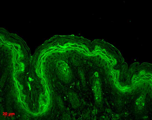
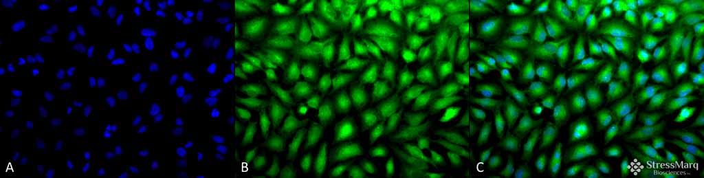
| Certificate of Analysis | A 1:1000 dilution of SPC-120 was sufficient for detection of ERK1/2 in 20 µg of HeLa cell lysate by ECL immunoblot analysis. |
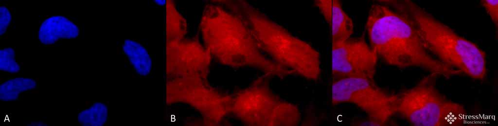
| Cellular Localization | Cytoplasm | Nucleus |

Thousands of laboratories across the world have published research that depended on the performance of antibodies from Abcepta to advance their research. Check out links to articles that cite our products in major peer-reviewed journals, organized by research category.
info@abcepta.com, and receive a free "I Love Antibodies" mug.

Provided below are standard protocols that you may find useful for product applications.
Background
The extracellular signal-regulated kinases 1 and 2 (ERK1 and ERK2), also called p44 and p42 MAP kinases, are members of the Mitogen Activated Protein Kinase (MAPK) family of proteins found in all eukaryotes. Because the 44 kDa ERK1 and the 42 kDa ERK2 are highly homologous and both function in the same protein kinase cascade, the two proteins are often referred to collectively as ERK1/2 or p44/p42 MAP kinase (1). They are both located in the cytosol and mitochondria (2). While the role of cytosol ERK1/2 is well studied and involved in multiple cellular functions (2), the role of mitochondrial ERK1/2 remains poorly understood. Both ERK 1 and 2 are activated by MEK1 or MEK2, by dual phosphorylation of a threonine and tyrosine residue in the activation loop (TEY motif) (1, 3). Either phosphorylation alone can induce an electrophoretic mobility shift, but both are required for activation of the kinase. This dual phosphorylation is efficiently detected by phosphorylation state-specific antibody directed to the pTEpY motif. Once activated, MAP kinases phosphorylate a broad spectrum of substrates, including cytoskeletal proteins, translation regulators, transcription factors, and the Rsk family of protein kinases (4). ERK1/2 activation is generally thought to confer a survival advantage to cells (5); however there is increasing evidence that suggests that the activation of ERK1/2 also contributes to cell death under certain conditions (5). ERK1/2 also is activated in neuronal and renal epithelial cells upon exposure to oxidative stress and toxicants or deprivation of growth factors, and inhibition of the ERK pathway blocks apoptosis (5).
References
1. Boulton TG. et al. (1991) Biochemistry. 30(1):278-86.
2. Yoon S., and Seger R. (2006) Growth Factors 24:21-44.
3. Wolf G. (2005) Antioxid Redox Signal 7:1337-1345.
4. Chuerland D., Marmor G., Shainskaya A. and Seger R. (2008) J Biol Chem. Epub: http://www.jbc.org/cgi/doi/10.1074/jbc.M709030200
5. Zhuang S., and Schnellmann R.G. (2006) J Pharmacol Exp Ther 319:991-997.
If you have used an Abcepta product and would like to share how it has performed, please click on the "Submit Review" button and provide the requested information. Our staff will examine and post your review and contact you if needed.
If you have any additional inquiries please email technical services at tech@abcepta.com.
Ordering Information
Other Products
Shipping Information
