


 Foundational characteristics of cancer include proliferation, angiogenesis, migration, evasion of apoptosis, and cellular immortality. Find key markers for these cellular processes and antibodies to detect them.
 The SUMOplot™ Analysis Program predicts and scores sumoylation sites in your protein. SUMOylation is a post-translational modification involved in various cellular processes, such as nuclear-cytosolic transport, transcriptional regulation, apoptosis, protein stability, response to stress, and progression through the cell cycle.
 The Autophagy Receptor Motif Plotter predicts and scores autophagy receptor binding sites in your protein. Identifying proteins connected to this pathway is critical to understanding the role of autophagy in physiological as well as pathological processes such as development, differentiation, neurodegenerative diseases, stress, infection, and cancer.
Erk1/2 Antibody
Rabbit Anti-Rat Erk1/2 Polyclonal
- SPECIFICATION
- CITATIONS
- PROTOCOLS
- BACKGROUND

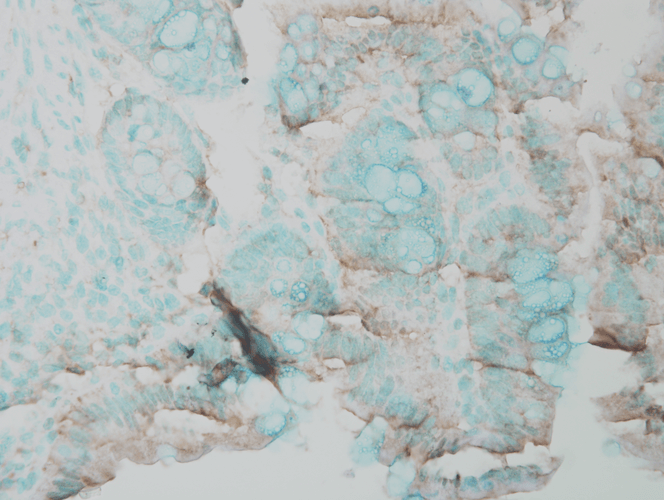
Application
| WB, IHC |
|---|---|
| Primary Accession | P21708 |
| Host | Rabbit |
| Reactivity | Human, Mouse, Rat, Chicken, Bovine, Drosophila, Xenopus, Sheep |
| Clonality | Polyclonal |
| Description | Rabbit Anti-Rat Erk1/2 Polyclonal |
| Target/Specificity | ERK1 |
| Other Names | ERK1 Antibody, ERK2 Antibody, ERT1 Antibody, ERT2 Antibody, MAP kinase1 Antibody, MAP kinase2 Antibody, MAPK1 Antibody, MAPK2 Antibody, MAPK3 Antibody, p38 Antibody, p40 Antibody, p41 Antibody, p41mapk Antibody, p42 MAPK Antibody, p44 ERK1 Antibody, p44 MAPK Antibody, PRKM1 PRKM2 Antibody, PRKM3 Antibody |
| Immunogen | A 35 residue synthetic peptide, corresponding to Rat Erk1 MAP kinase with the CGG spacer group added and the peptide coupled to KLH. |
| Purification | Peptide Affinity Purified |
| Storage | 1 mg/ml |
| Storage Buffer | PBS pH7.4, 50% glycerol, 0.09% sodium azide *Storage buffer may change when conjugated |
| Shipping Temperature | -20ºC |
| Certificate of Analysis | Blue Ice or 4ºC |
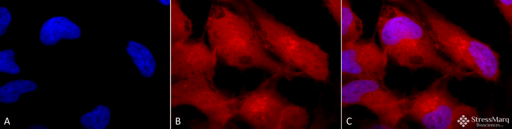
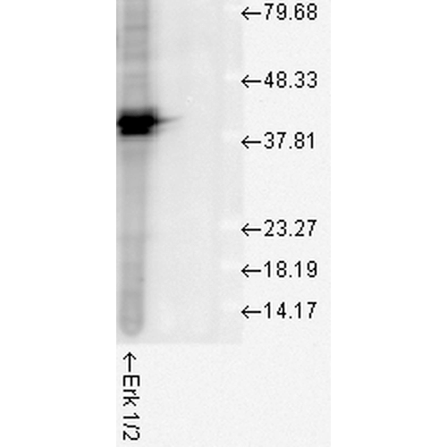
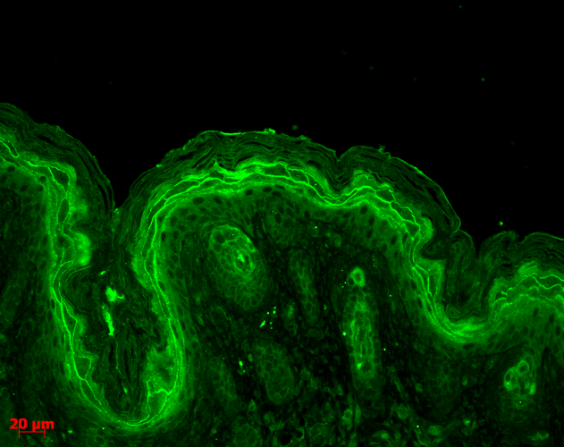
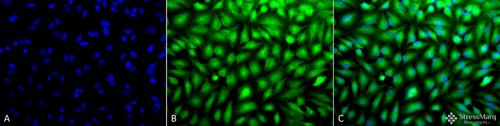
| Cellular Localization | A 1:1000 dilution of ASM10707 was sufficient for detection of ERK1/2 in 20 µg of HeLa cell lysate by ECL immunoblot analysis. |
Research Areas
Citations (0)

Thousands of laboratories across the world have published research that depended on the performance of antibodies from Abcepta to advance their research. Check out links to articles that cite our products in major peer-reviewed journals, organized by research category.
Submit your citation using an Abcepta antibody to
info@abcepta.com, and receive a free "I Love Antibodies" mug.
info@abcepta.com, and receive a free "I Love Antibodies" mug.

Application Protocols
Provided below are standard protocols that you may find useful for product applications.
Abcepta welcomes feedback from its customers.
If you have used an Abcepta product and would like to share how it has performed, please click on the "Submit Review" button and provide the requested information. Our staff will examine and post your review and contact you if needed.
If you have any additional inquiries please email technical services at tech@abcepta.com.
Cat# ASM10707
Ordering Information
United States
AlbaniaAustraliaAustriaBelgiumBosnia & HerzegovinaBrazilBulgariaCanadaCentral AmericaChinaCroatiaCyprusCzech RepublicDenmarkEstoniaFinlandFranceGermanyGreeceHong KongHungaryIcelandIndiaIndonesiaIrelandIsraelItalyJapanLatviaLithuaniaLuxembourgMacedoniaMalaysiaMaltaMexicoNetherlandsNew ZealandNorwayPakistanPolandPortugalRomaniaSerbiaSingaporeSlovakiaSloveniaSouth AfricaSouth KoreaSpainSwedenSwitzerlandTaiwanTurkeyUnited KingdomUnited StatesVietnamWorldwideOthers
USA Headquarters
(888) 735-7227 / (858) 622-0099 or (858) 875-1900
Other Products
Shipping Information
Domestic orders (in stock items)
Shipped out the same day. Orders placed after 1 PM (PST) will ship out the next business day.
International orders
Contact your local distributors
