


 Foundational characteristics of cancer include proliferation, angiogenesis, migration, evasion of apoptosis, and cellular immortality. Find key markers for these cellular processes and antibodies to detect them.
 The SUMOplot™ Analysis Program predicts and scores sumoylation sites in your protein. SUMOylation is a post-translational modification involved in various cellular processes, such as nuclear-cytosolic transport, transcriptional regulation, apoptosis, protein stability, response to stress, and progression through the cell cycle.
 The Autophagy Receptor Motif Plotter predicts and scores autophagy receptor binding sites in your protein. Identifying proteins connected to this pathway is critical to understanding the role of autophagy in physiological as well as pathological processes such as development, differentiation, neurodegenerative diseases, stress, infection, and cancer.
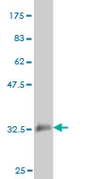
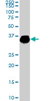
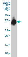
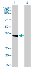
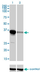
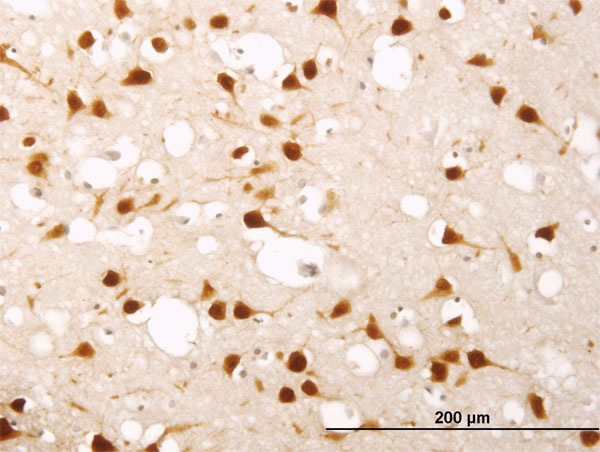
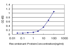
ELAVL4 Antibody (monoclonal) (M01)
Mouse monoclonal antibody raised against a partial recombinant ELAVL4.
- SPECIFICATION
- CITATIONS
- PROTOCOLS
- BACKGROUND

Application
| WB, IHC, E |
|---|---|
| Primary Accession | P26378 |
| Other Accession | NM_021952 |
| Reactivity | Human, Rat |
| Host | mouse |
| Clonality | Monoclonal |
| Isotype | IgG1 Kappa |
| Clone Names | 6B9 |
| Calculated MW | 42398 Da |
| Gene ID | 1996 |
|---|---|
| Other Names | ELAV-like protein 4, Hu-antigen D, HuD, Paraneoplastic encephalomyelitis antigen HuD, ELAVL4, HUD, PNEM |
| Target/Specificity | ELAVL4 (NP_068771, 312 a.a. ~ 380 a.a) partial recombinant protein with GST tag. MW of the GST tag alone is 26 KDa. |
| Dilution | WB~~1:500~1000 IHC~~1:100~500 E~~N/A |
| Format | Clear, colorless solution in phosphate buffered saline, pH 7.2 . |
| Storage | Store at -20°C or lower. Aliquot to avoid repeated freezing and thawing. |
| Precautions | ELAVL4 Antibody (monoclonal) (M01) is for research use only and not for use in diagnostic or therapeutic procedures. |

Thousands of laboratories across the world have published research that depended on the performance of antibodies from Abcepta to advance their research. Check out links to articles that cite our products in major peer-reviewed journals, organized by research category.
info@abcepta.com, and receive a free "I Love Antibodies" mug.

Provided below are standard protocols that you may find useful for product applications.
References
Molecular analysis of the HuD gene in neuroendocrine lung cancers. D'Alessandro V, et al. Lung Cancer, 2010 Jan. PMID 19410329.No evidence for the presence of HuD-specific T cells in the cerebrospinal fluid of patients with Hu-associated paraneoplastic neurological syndromes. de Beukelaar JW, et al. J Neurol, 2009 Feb. PMID 19252764.Molecular detection of neuron-specific ELAV-like-positive cells in the peripheral blood of patients with small-cell lung cancer. D'Alessandro V, et al. Cell Oncol, 2008. PMID 18607064.Replication of association between ELAVL4 and Parkinson disease: the GenePD study. DeStefano AL, et al. Hum Genet, 2008 Aug. PMID 18587682.Alterations in mossy fiber physiology and GAP-43 expression and function in transgenic mice overexpressing HuD. Tanner DC, et al. Hippocampus, 2008. PMID 18493953.
If you have used an Abcepta product and would like to share how it has performed, please click on the "Submit Review" button and provide the requested information. Our staff will examine and post your review and contact you if needed.
If you have any additional inquiries please email technical services at tech@abcepta.com.
Ordering Information
Other Products
Shipping Information
