


 Foundational characteristics of cancer include proliferation, angiogenesis, migration, evasion of apoptosis, and cellular immortality. Find key markers for these cellular processes and antibodies to detect them.
 The SUMOplot™ Analysis Program predicts and scores sumoylation sites in your protein. SUMOylation is a post-translational modification involved in various cellular processes, such as nuclear-cytosolic transport, transcriptional regulation, apoptosis, protein stability, response to stress, and progression through the cell cycle.
 The Autophagy Receptor Motif Plotter predicts and scores autophagy receptor binding sites in your protein. Identifying proteins connected to this pathway is critical to understanding the role of autophagy in physiological as well as pathological processes such as development, differentiation, neurodegenerative diseases, stress, infection, and cancer.
Anti-DR5 Picoband Antibody
- SPECIFICATION
- CITATIONS
- PROTOCOLS
- BACKGROUND

Application
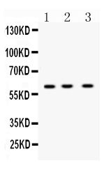
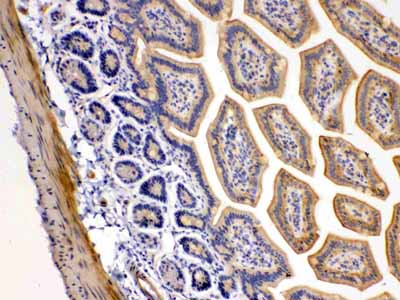
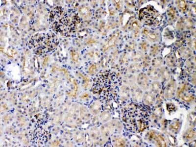
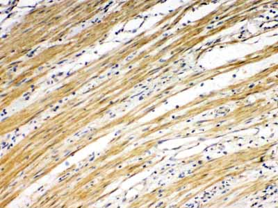
| WB, IHC-P |
|---|---|
| Primary Accession | O14763 |
| Host | Rabbit |
| Reactivity | Human, Mouse, Rat |
| Clonality | Polyclonal |
| Format | Lyophilized |
| Description | Rabbit IgG polyclonal antibody for Tumor necrosis factor receptor superfamily member 10B(TNFRSF10B) detection. Tested with WB, IHC-P in Human;Mouse;Rat. |
| Reconstitution | Add 0.2ml of distilled water will yield a concentration of 500ug/ml. |
| Gene ID | 8795 |
|---|---|
| Other Names | Tumor necrosis factor receptor superfamily member 10B, Death receptor 5, TNF-related apoptosis-inducing ligand receptor 2, TRAIL receptor 2, TRAIL-R2, CD262, TNFRSF10B, DR5, KILLER, TRAILR2, TRICK2, ZTNFR9 |
| Calculated MW | 47878 MW KDa |
| Application Details | Immunohistochemistry(Paraffin-embedded Section), 0.5-1 µg/ml, Human, Mouse, Rat, By Heat Western blot, 0.1-0.5 µg/ml, Human, Mouse, Rat |
| Subcellular Localization | Membrane; Single-pass type I membrane protein. |
| Tissue Specificity | Widely expressed in adult and fetal tissues; very highly expressed in tumor cell lines such as HeLaS3, K-562, HL-60, SW480, A-549 and G-361; highly expressed in heart, peripheral blood lymphocytes, liver, pancreas, spleen, thymus, prostate, ovary, uterus, placenta, testis, esophagus, stomach and throughout the intestinal tract; not detectable in brain. |
| Protein Name | Tumor necrosis factor receptor superfamily member 10B |
| Contents | Each vial contains 5mg BSA, 0.9mg NaCl, 0.2mg Na2HPO4, 0.05mg NaN3. |
| Immunogen | E.coli-derived human DR5 recombinant protein (Position: K233-S440). Human DR5 shares 48.4% amino acid (aa) sequence identity with mouse DR5. |
| Purification | Immunogen affinity purified. |
| Cross Reactivity | No cross reactivity with other proteins |
| Storage | At -20˚C for one year. After r˚Constitution, at 4˚C for one month. It˚Can also be aliquotted and stored frozen at -20˚C for a longer time.Avoid repeated freezing and thawing. |
| Name | TNFRSF10B |
|---|---|
| Synonyms | DR5, KILLER, TRAILR2, TRICK2, ZTNFR9 |
| Function | Receptor for the cytotoxic ligand TNFSF10/TRAIL (PubMed:10549288). The adapter molecule FADD recruits caspase-8 to the activated receptor. The resulting death-inducing signaling complex (DISC) performs caspase-8 proteolytic activation which initiates the subsequent cascade of caspases (aspartate-specific cysteine proteases) mediating apoptosis. Promotes the activation of NF-kappa-B. Essential for ER stress-induced apoptosis. |
| Cellular Location | Membrane; Single-pass type I membrane protein. |
| Tissue Location | Widely expressed in adult and fetal tissues; very highly expressed in tumor cell lines such as HeLaS3, K-562, HL-60, SW480, A-549 and G-361; highly expressed in heart, peripheral blood lymphocytes, liver, pancreas, spleen, thymus, prostate, ovary, uterus, placenta, testis, esophagus, stomach and throughout the intestinal tract; not detectable in brain |

Thousands of laboratories across the world have published research that depended on the performance of antibodies from Abcepta to advance their research. Check out links to articles that cite our products in major peer-reviewed journals, organized by research category.
info@abcepta.com, and receive a free "I Love Antibodies" mug.

Provided below are standard protocols that you may find useful for product applications.
Background
TNFRSF10B(Tumor necrosis factor receptor superfamily, member 10b) is a human gene. It is also known as DR5, CD262, KILLER, TRICK2, TRICKB, ZTNFR9, TRAILR2, TRICK2A, TRICK2B, TRAIL-R2, KILLER/DR5. The protein encoded by this gene is a member of the TNF-receptor superfamily, and contains an intracellular death domain. This receptor can be activated by tumor necrosis factor-related apoptosis inducing ligand (TNFSF10/TRAIL/APO-2L), and transduces apoptosis signal. Mice have a homologous gene, tnfrsf10b that has been essential in the elucidation of the function of this gene in humans. Studies with FADD-deficient mice suggested that FADD, a death domain containing adaptor protein, is required for the apoptosis mediated by this protein.By analysis of radiation hybrid panels, this gene is mapped to chromosome 8p22-p21. Northern blot analysis indicated that TRAILR2 was expressed as a 4.4-kb mRNA in all tissues tested, with the highest levels of expression in peripheral blood lymphocytes, spleen, and ovary.
If you have used an Abcepta product and would like to share how it has performed, please click on the "Submit Review" button and provide the requested information. Our staff will examine and post your review and contact you if needed.
If you have any additional inquiries please email technical services at tech@abcepta.com.
Ordering Information
Other Products
Shipping Information
