


 Foundational characteristics of cancer include proliferation, angiogenesis, migration, evasion of apoptosis, and cellular immortality. Find key markers for these cellular processes and antibodies to detect them.
 The SUMOplot™ Analysis Program predicts and scores sumoylation sites in your protein. SUMOylation is a post-translational modification involved in various cellular processes, such as nuclear-cytosolic transport, transcriptional regulation, apoptosis, protein stability, response to stress, and progression through the cell cycle.
 The Autophagy Receptor Motif Plotter predicts and scores autophagy receptor binding sites in your protein. Identifying proteins connected to this pathway is critical to understanding the role of autophagy in physiological as well as pathological processes such as development, differentiation, neurodegenerative diseases, stress, infection, and cancer.
Anti-Periplakin Picoband Antibody
- SPECIFICATION
- CITATIONS
- PROTOCOLS
- BACKGROUND

Application
| WB, IHC-P |
|---|---|
| Primary Accession | O60437 |
| Host | Rabbit |
| Reactivity | Human, Mouse, Rat |
| Clonality | Polyclonal |
| Format | Lyophilized |
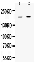
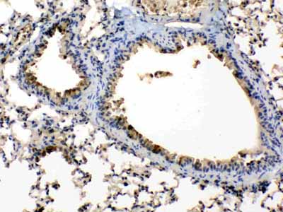
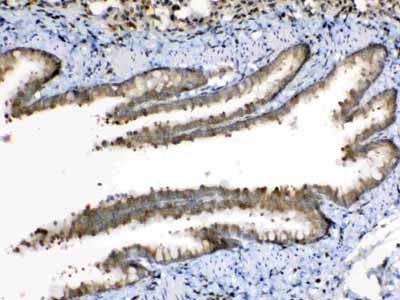
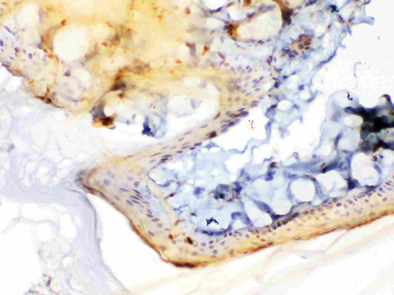
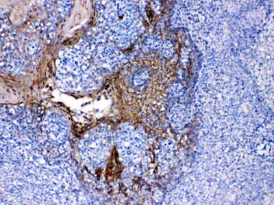
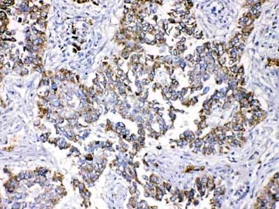
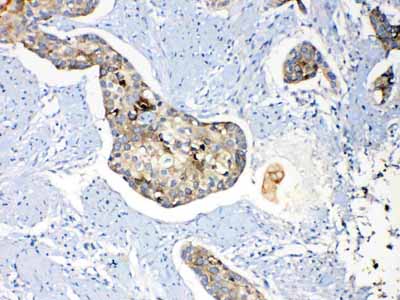
| Description | Rabbit IgG polyclonal antibody for Periplakin(PPL) detection. Tested with WB, IHC-P in Human;Mouse;Rat. |
| Reconstitution | Add 0.2ml of distilled water will yield a concentration of 500ug/ml. |
| Gene ID | 5493 |
|---|---|
| Other Names | Periplakin, 190 kDa paraneoplastic pemphigus antigen, 195 kDa cornified envelope precursor protein, PPL, KIAA0568 |
| Calculated MW | 204747 MW KDa |
| Application Details | Immunohistochemistry(Paraffin-embedded Section), 0.5-1 µg/ml, Human, Mouse, Rat, By Heat Western blot, 0.1-0.5 µg/ml, Human, Rat |
| Subcellular Localization | Cell junction, desmosome . Cytoplasm, cytoskeleton . Cell membrane . Nucleus . Mitochondrion . Associated with desmosomes and intermediate filaments. |
| Tissue Specificity | Expressed in stratified squamous epithelia and in some other epithelia. . |
| Protein Name | Periplakin |
| Contents | Each vial contains 5mg BSA, 0.9mg NaCl, 0.2mg Na2HPO4, 0.05mg NaN3. |
| Immunogen | A synthetic peptide corresponding to a sequence at the C-terminus of human Periplakin (1664-1701aa DTGRELSPEEAHRAGLIDWNMFVKLRSQECDWEEISVK), different from the related mouse sequence by one amino acid. |
| Purification | Immunogen affinity purified. |
| Cross Reactivity | No cross reactivity with other proteins |
| Storage | At -20˚C for one year. After r˚Constitution, at 4˚C for one month. It˚Can also be aliquotted and stored frozen at -20˚C for a longer time.Avoid repeated freezing and thawing. |
| Name | PPL |
|---|---|
| Synonyms | KIAA0568 |
| Function | Component of the cornified envelope of keratinocytes. May link the cornified envelope to desmosomes and intermediate filaments. May act as a localization signal in PKB/AKT-mediated signaling. |
| Cellular Location | Cell junction, desmosome. Cytoplasm, cytoskeleton. Cell membrane {ECO:0000250|UniProtKB:Q9R269}. Cytoplasm {ECO:0000250|UniProtKB:Q9R269} |
| Tissue Location | Expressed in stratified squamous epithelia and in some other epithelia. |

Thousands of laboratories across the world have published research that depended on the performance of antibodies from Abcepta to advance their research. Check out links to articles that cite our products in major peer-reviewed journals, organized by research category.
info@abcepta.com, and receive a free "I Love Antibodies" mug.

Provided below are standard protocols that you may find useful for product applications.
Background
Periplakin is a protein that in humans is encoded by the PPL gene. The protein encoded by this gene is a component of desmosomes and of the epidermal cornified envelope in keratinocytes. The N-terminal domain of this protein interacts with the plasma membrane and its C-terminus interacts with intermediate filaments. Through its rod domain, this protein forms complexes with envoplakin. This protein may serve as a link between the cornified envelope and desmosomes as well as intermediate filaments. AKT1/PKB, a protein kinase mediating a variety of cell growth and survival signaling processes, is reported to interact with this protein, suggesting a possible role for this protein as a localization signal in AKT1-mediated signaling.
If you have used an Abcepta product and would like to share how it has performed, please click on the "Submit Review" button and provide the requested information. Our staff will examine and post your review and contact you if needed.
If you have any additional inquiries please email technical services at tech@abcepta.com.
Ordering Information
Shipping Information
