


 Foundational characteristics of cancer include proliferation, angiogenesis, migration, evasion of apoptosis, and cellular immortality. Find key markers for these cellular processes and antibodies to detect them.
 The SUMOplot™ Analysis Program predicts and scores sumoylation sites in your protein. SUMOylation is a post-translational modification involved in various cellular processes, such as nuclear-cytosolic transport, transcriptional regulation, apoptosis, protein stability, response to stress, and progression through the cell cycle.
 The Autophagy Receptor Motif Plotter predicts and scores autophagy receptor binding sites in your protein. Identifying proteins connected to this pathway is critical to understanding the role of autophagy in physiological as well as pathological processes such as development, differentiation, neurodegenerative diseases, stress, infection, and cancer.
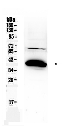
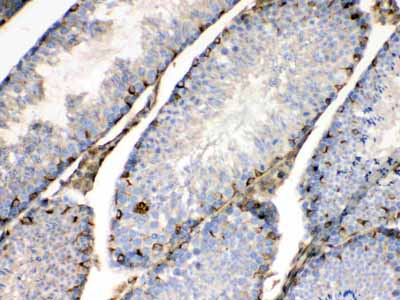
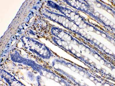
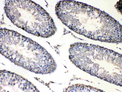
Anti-Bag1 Picoband Antibody
- SPECIFICATION
- CITATIONS
- PROTOCOLS
- BACKGROUND

Application
| WB, IHC-P |
|---|---|
| Primary Accession | Q99933 |
| Host | Rabbit |
| Reactivity | Human, Mouse, Rat |
| Clonality | Polyclonal |
| Format | Lyophilized |
| Description | Rabbit IgG polyclonal antibody for BAG family molecular chaperone regulator 1(BAG1) detection. Tested with WB, IHC-P in Human;Mouse;Rat. |
| Reconstitution | Add 0.2ml of distilled water will yield a concentration of 500ug/ml. |
| Gene ID | 573 |
|---|---|
| Other Names | BAG family molecular chaperone regulator 1, BAG-1, Bcl-2-associated athanogene 1, BAG1, HAP |
| Calculated MW | 38779 MW KDa |
| Application Details | Immunohistochemistry(Paraffin-embedded Section), 0.5-1 µg/ml, Mouse, Rat, Human, By Heat Western blot, 0.1-0.5 µg/ml, Rat, Human |
| Subcellular Localization | Isoform 1: Nucleus. Cytoplasm. Isoform 1 localizes predominantly to the nucleus. |
| Tissue Specificity | Isoform 4 is the most abundantly expressed isoform. It is ubiquitously expressed throughout most tissues, except the liver, colon, breast and uterine myometrium. Isoform 1 is expressed in the ovary and testis. Isoform 4 is expressed in several types of tumor cell lines, and at consistently high levels in leukemia and lymphoma cell lines. Isoform 1 is expressed in the prostate, breast and leukemia cell lines. Isoform 3 is the least abundant isoform in tumor cell lines (at protein level). . |
| Protein Name | BAG family molecular chaperone regulator 1 |
| Contents | Each vial contains 5mg BSA, 0.9mg NaCl, 0.2mg Na2HPO4, 0.05mg NaN3. |
| Immunogen | A synthetic peptide corresponding to a sequence at the C-terminus of human Bag1 (323-345aa DTVEQNICQETERLQSTNFALAE), different from the related mouse sequence by two amino acids, and from the related rat sequence by three amino acids. |
| Purification | Immunogen affinity purified. |
| Cross Reactivity | No cross reactivity with other proteins |
| Storage | At -20˚C for one year. After r˚Constitution, at 4˚C for one month. It˚Can also be aliquotted and stored frozen at -20˚C for a longer time.Avoid repeated freezing and thawing. |
| Name | BAG1 |
|---|---|
| Synonyms | HAP |
| Function | Co-chaperone for HSP70 and HSC70 chaperone proteins. Acts as a nucleotide-exchange factor (NEF) promoting the release of ADP from the HSP70 and HSC70 proteins thereby triggering client/substrate protein release. Nucleotide release is mediated via its binding to the nucleotide-binding domain (NBD) of HSPA8/HSC70 where as the substrate release is mediated via its binding to the substrate-binding domain (SBD) of HSPA8/HSC70 (PubMed:24318877, PubMed:27474739, PubMed:9873016). Inhibits the pro-apoptotic function of PPP1R15A, and has anti-apoptotic activity (PubMed:12724406). Markedly increases the anti-cell death function of BCL2 induced by various stimuli (PubMed:9305631). Involved in the STUB1-mediated proteasomal degradation of ESR1 in response to age-related circulating estradiol (17-beta-estradiol/E2) decline, thereby promotes neuronal apoptosis in response to ischemic reperfusion injury (By similarity). |
| Cellular Location | [Isoform 1]: Nucleus. Cytoplasm. Note=Isoform 1 localizes predominantly to the nucleus [Isoform 4]: Cytoplasm. Nucleus. Note=Isoform 4 localizes predominantly to the cytoplasm. The cellular background in which it is expressed can influence whether it resides primarily in the cytoplasm or is also found in the nucleus. In the presence of BCL2, localizes to intracellular membranes (what appears to be the nuclear envelope and perinuclear membranes) as well as punctate cytosolic structures suggestive of mitochondria |
| Tissue Location | Isoform 4 is the most abundantly expressed isoform. It is ubiquitously expressed throughout most tissues, except the liver, colon, breast and uterine myometrium. Isoform 1 is expressed in the ovary and testis. Isoform 4 is expressed in several types of tumor cell lines, and at consistently high levels in leukemia and lymphoma cell lines. Isoform 1 is expressed in the prostate, breast and leukemia cell lines. Isoform 3 is the least abundant isoform in tumor cell lines (at protein level). |

Thousands of laboratories across the world have published research that depended on the performance of antibodies from Abcepta to advance their research. Check out links to articles that cite our products in major peer-reviewed journals, organized by research category.
info@abcepta.com, and receive a free "I Love Antibodies" mug.

Provided below are standard protocols that you may find useful for product applications.
Background
BAG family molecular chaperone regulator 1 (BAG1) is a protein that in humans is encoded by the BAG1 gene. Human BAG1 is mapped to chromosome 9p12, a region associated with hereditary disorders that may involve developmental dysregulation of programmed cell death. The Bag1 protein is rich in glutamic acid residues. Its deduced 274-amino acid protein has a calculated molecular mass of 31 KD. Being the BCL-2-associated athanogene, Bag1 enhances the anti-apoptotic effects of BCL2 and represents a link between growth factor receptors and anti-apoptotic mechanisms.
If you have used an Abcepta product and would like to share how it has performed, please click on the "Submit Review" button and provide the requested information. Our staff will examine and post your review and contact you if needed.
If you have any additional inquiries please email technical services at tech@abcepta.com.
Ordering Information
Other Products
Shipping Information
