


 Foundational characteristics of cancer include proliferation, angiogenesis, migration, evasion of apoptosis, and cellular immortality. Find key markers for these cellular processes and antibodies to detect them.
 The SUMOplot™ Analysis Program predicts and scores sumoylation sites in your protein. SUMOylation is a post-translational modification involved in various cellular processes, such as nuclear-cytosolic transport, transcriptional regulation, apoptosis, protein stability, response to stress, and progression through the cell cycle.
 The Autophagy Receptor Motif Plotter predicts and scores autophagy receptor binding sites in your protein. Identifying proteins connected to this pathway is critical to understanding the role of autophagy in physiological as well as pathological processes such as development, differentiation, neurodegenerative diseases, stress, infection, and cancer.
Anti-BRMS1 Picoband Antibody
- SPECIFICATION
- CITATIONS
- PROTOCOLS
- BACKGROUND

Application
| WB |
|---|---|
| Primary Accession | Q9HCU9 |
| Host | Rabbit |
| Reactivity | Human, Mouse, Rat |
| Clonality | Polyclonal |
| Format | Lyophilized |
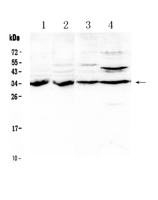
| Description | Rabbit IgG polyclonal antibody for Breast cancer metastasis-suppressor 1(BRMS1) detection. Tested with WB in Human;Mouse;Rat. |
| Reconstitution | Add 0.2ml of distilled water will yield a concentration of 500ug/ml. |
| Gene ID | 25855 |
|---|---|
| Other Names | Breast cancer metastasis-suppressor 1, BRMS1 |
| Calculated MW | 28461 MW KDa |
| Application Details | Western blot, 0.1-0.5 µg/ml, Human, Mouse, Rat |
| Subcellular Localization | Nucleus. Cytoplasm. Predominantly nuclear. |
| Tissue Specificity | Expression levels are higher in term placentas than in early placentas. Low levels of expression observed in normal pregnancies and in molar pregnancies. . |
| Protein Name | Breast cancer metastasis-suppressor 1 |
| Contents | Each vial contains 5mg BSA, 0.9mg NaCl, 0.2mg Na2HPO4, 0.05mg NaN3. |
| Immunogen | E.coli-derived human BRMS1 recombinant protein (Position: D52-D244). Human BRMS1 shares 93.6% and 96% amino acid (aa) sequence identity with mouse and rat BRMS1, respectively. |
| Purification | Immunogen affinity purified. |
| Cross Reactivity | No cross reactivity with other proteins. |
| Storage | At -20˚C for one year. After r˚Constitution, at 4˚C for one month. It˚Can also be aliquotted and stored frozen at -20˚C for a longer time.Avoid repeated freezing and thawing. |
| Name | BRMS1 |
|---|---|
| Function | Transcriptional repressor. Down-regulates transcription activation by NF-kappa-B by promoting the deacetylation of RELA at 'Lys-310'. Promotes HDAC1 binding to promoter regions. Down-regulates expression of anti-apoptotic genes that are controlled by NF-kappa-B. Promotes apoptosis in cells that have inadequate adherence to a substrate, a process called anoikis, and may thereby inhibit metastasis. May be a mediator of metastasis suppression in breast carcinoma. |
| Cellular Location | Nucleus. Cytoplasm. Note=Predominantly nuclear. |
| Tissue Location | Expression levels are higher in term placentas than in early placentas. Low levels of expression observed in normal pregnancies and in molar pregnancies. |

Thousands of laboratories across the world have published research that depended on the performance of antibodies from Abcepta to advance their research. Check out links to articles that cite our products in major peer-reviewed journals, organized by research category.
info@abcepta.com, and receive a free "I Love Antibodies" mug.

Provided below are standard protocols that you may find useful for product applications.
Background
Breast cancer metastasis-suppressor 1 is a protein that in humans is encoded by the BRMS1 gene. This gene reduces the metastatic potential, but not the tumorogenicity, of human breast cancer and melanoma cell lines. The protein encoded by this gene localizes primarily to the nucleus and is a component of the mSin3a family of histone deacetylase complexes (HDAC). The protein contains two coiled-coil motifs and several imperfect leucine zipper motifs. Alternative splicing results in two transcript variants encoding different isoforms.
If you have used an Abcepta product and would like to share how it has performed, please click on the "Submit Review" button and provide the requested information. Our staff will examine and post your review and contact you if needed.
If you have any additional inquiries please email technical services at tech@abcepta.com.
Ordering Information
Other Products
Shipping Information
