


 Foundational characteristics of cancer include proliferation, angiogenesis, migration, evasion of apoptosis, and cellular immortality. Find key markers for these cellular processes and antibodies to detect them.
 The SUMOplot™ Analysis Program predicts and scores sumoylation sites in your protein. SUMOylation is a post-translational modification involved in various cellular processes, such as nuclear-cytosolic transport, transcriptional regulation, apoptosis, protein stability, response to stress, and progression through the cell cycle.
 The Autophagy Receptor Motif Plotter predicts and scores autophagy receptor binding sites in your protein. Identifying proteins connected to this pathway is critical to understanding the role of autophagy in physiological as well as pathological processes such as development, differentiation, neurodegenerative diseases, stress, infection, and cancer.
Anti-Musashi 1/Msi1 Picoband Antibody
- SPECIFICATION
- CITATIONS
- PROTOCOLS
- BACKGROUND

Application
| WB, IHC-P |
|---|---|
| Primary Accession | O43347 |
| Host | Rabbit |
| Reactivity | Human, Mouse, Rat |
| Clonality | Polyclonal |
| Format | Lyophilized |
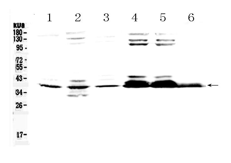
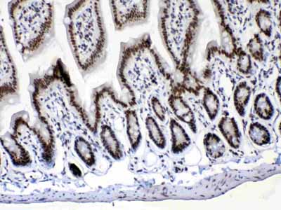
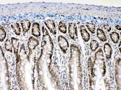
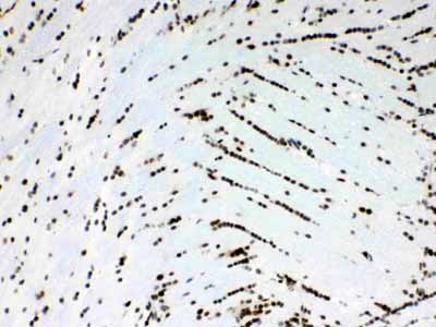
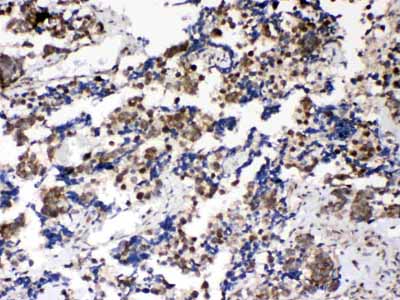
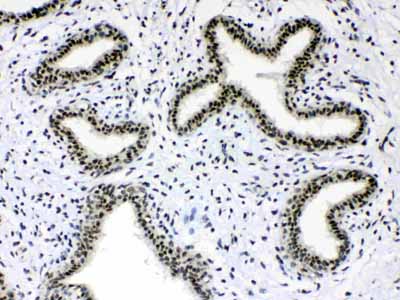
| Description | Rabbit IgG polyclonal antibody for RNA-binding protein Musashi homolog 1(MSI1) detection. Tested with WB, IHC-P in Human;Mouse;Rat. |
| Reconstitution | Add 0.2ml of distilled water will yield a concentration of 500ug/ml. |
| Gene ID | 4440 |
|---|---|
| Other Names | RNA-binding protein Musashi homolog 1, Musashi-1, MSI1 |
| Calculated MW | 39125 MW KDa |
| Application Details | Immunohistochemistry(Paraffin-embedded Section), 0.5-1 µg/ml, Human, Mouse, Rat, By Heat Western blot, 0.1-0.5 µg/ml, Human, Mouse, Rat |
| Subcellular Localization | Cytoplasm . Nucleus . |
| Tissue Specificity | Detected in fetal kidney, brain, liver and lung, and in adult brain and pancreas. Detected in hepatoma cell lines. . |
| Protein Name | RNA-binding protein Musashi homolog 1 |
| Contents | Each vial contains 5mg BSA, 0.9mg NaCl, 0.2mg Na2HPO4, 0.05mg NaN3. |
| Immunogen | A synthetic peptide corresponding to a sequence at the N-terminus of human Musashi 1/Msi1 (21-54aa KMFIGGLSWQTTQEGLREYFGQFGEVKECLVMRD), identical to the related mouse and rat sequences. |
| Purification | Immunogen affinity purified. |
| Cross Reactivity | No cross reactivity with other proteins |
| Storage | At -20˚C for one year. After r˚Constitution, at 4˚C for one month. It˚Can also be aliquotted and stored frozen at -20˚C for a longer time.Avoid repeated freezing and thawing. |
| Name | MSI1 |
|---|---|
| Function | RNA binding protein that regulates the expression of target mRNAs at the translation level. Regulates expression of the NOTCH1 antagonist NUMB. Binds RNA containing the sequence 5'-GUUAGUUAGUUAGUU- 3' and other sequences containing the pattern 5'-[GA]U(1-3)AGU-3'. May play a role in the proliferation and maintenance of stem cells in the central nervous system (By similarity). |
| Cellular Location | Cytoplasm {ECO:0000250|UniProtKB:Q61474}. Nucleus {ECO:0000250|UniProtKB:Q61474} |
| Tissue Location | Detected in fetal kidney, brain, liver and lung, and in adult brain and pancreas. Detected in hepatoma cell lines |

Thousands of laboratories across the world have published research that depended on the performance of antibodies from Abcepta to advance their research. Check out links to articles that cite our products in major peer-reviewed journals, organized by research category.
info@abcepta.com, and receive a free "I Love Antibodies" mug.

Provided below are standard protocols that you may find useful for product applications.
Background
RNA-binding protein Musashi homolog 1 is a protein that in humans is encoded by the MSI1 gene. This gene encodes a protein containing two conserved tandem RNA recognition motifs. Similar proteins in other species function as RNA-binding proteins and play central roles in posttranscriptional gene regulation. Expression of this gene has been correlated with the grade of the malignancy and proliferative activity in gliomas and melanomas. A pseudogene for this gene is located on chromosome 11q13.
If you have used an Abcepta product and would like to share how it has performed, please click on the "Submit Review" button and provide the requested information. Our staff will examine and post your review and contact you if needed.
If you have any additional inquiries please email technical services at tech@abcepta.com.
Ordering Information
Other Products
Shipping Information
