


 Foundational characteristics of cancer include proliferation, angiogenesis, migration, evasion of apoptosis, and cellular immortality. Find key markers for these cellular processes and antibodies to detect them.
 The SUMOplot™ Analysis Program predicts and scores sumoylation sites in your protein. SUMOylation is a post-translational modification involved in various cellular processes, such as nuclear-cytosolic transport, transcriptional regulation, apoptosis, protein stability, response to stress, and progression through the cell cycle.
 The Autophagy Receptor Motif Plotter predicts and scores autophagy receptor binding sites in your protein. Identifying proteins connected to this pathway is critical to understanding the role of autophagy in physiological as well as pathological processes such as development, differentiation, neurodegenerative diseases, stress, infection, and cancer.
Anti-AP2M1 Picoband Antibody
- SPECIFICATION
- CITATIONS
- PROTOCOLS
- BACKGROUND

Application
| WB |
|---|---|
| Primary Accession | Q96CW1 |
| Host | Rabbit |
| Reactivity | Human, Mouse, Rat |
| Clonality | Polyclonal |
| Format | Lyophilized |
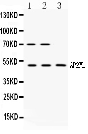
| Description | Rabbit IgG polyclonal antibody for AP-2 complex subunit mu(AP2M1) detection. Tested with WB in Human;Mouse;Rat. |
| Reconstitution | Add 0.2ml of distilled water will yield a concentration of 500ug/ml. |
| Gene ID | 1173 |
|---|---|
| Other Names | AP-2 complex subunit mu, AP-2 mu chain, Adaptin-mu2, Adaptor protein complex AP-2 subunit mu, Adaptor-related protein complex 2 subunit mu, Clathrin assembly protein complex 2 mu medium chain, Clathrin coat assembly protein AP50, Clathrin coat-associated protein AP50, HA2 50 kDa subunit, Plasma membrane adaptor AP-2 50 kDa protein, AP2M1, CLAPM1, KIAA0109 |
| Calculated MW | 49655 MW KDa |
| Application Details | Western blot, 0.1-0.5 µg/ml, Human, Mouse, Rat |
| Subcellular Localization | Cell membrane. Membrane, coated pit; Peripheral membrane protein; Cytoplasmic side. AP-2 appears to be excluded from internalizing CCVs and to disengage from sites of endocytosis seconds before internalization of the nascent CCV. . |
| Protein Name | AP-2 complex subunit mu |
| Contents | Each vial contains 5mg BSA, 0.9mg NaCl, 0.2mg Na2HPO4, 0.05mg NaN3. |
| Immunogen | A synthetic peptide corresponding to a sequence at the C-terminus of human AP2M1 (399-435aa LKVRYLKVFEPKLNYSDHDVIKWVRYIGRSGIYETRC), identical to the related mouse and rat sequences. |
| Purification | Immunogen affinity purified. |
| Cross Reactivity | No cross reactivity with other proteins. |
| Storage | At -20˚C for one year. After r˚Constitution, at 4˚C for one month. It˚Can also be aliquotted and stored frozen at -20˚C for a longer time.Avoid repeated freezing and thawing. |
| Name | AP2M1 (HGNC:564) |
|---|---|
| Synonyms | CLAPM1, KIAA0109 |
| Function | Component of the adaptor protein complex 2 (AP-2) (PubMed:12694563, PubMed:12952941, PubMed:14745134, PubMed:14985334, PubMed:15473838, PubMed:31104773). Adaptor protein complexes function in protein transport via transport vesicles in different membrane traffic pathways (PubMed:12694563, PubMed:12952941, PubMed:14745134, PubMed:14985334, PubMed:15473838, PubMed:31104773). Adaptor protein complexes are vesicle coat components and appear to be involved in cargo selection and vesicle formation (PubMed:12694563, PubMed:12952941, PubMed:14745134, PubMed:14985334, PubMed:15473838, PubMed:31104773). AP-2 is involved in clathrin-dependent endocytosis in which cargo proteins are incorporated into vesicles surrounded by clathrin (clathrin-coated vesicles, CCVs) which are destined for fusion with the early endosome (PubMed:12694563, PubMed:12952941, PubMed:14745134, PubMed:14985334, PubMed:15473838, PubMed:31104773). The clathrin lattice serves as a mechanical scaffold but is itself unable to bind directly to membrane components (PubMed:12694563, PubMed:12952941, PubMed:14745134, PubMed:14985334, PubMed:15473838, PubMed:31104773). Clathrin-associated adaptor protein (AP) complexes which can bind directly to both the clathrin lattice and to the lipid and protein components of membranes are considered to be the major clathrin adaptors contributing the CCV formation (PubMed:12694563, PubMed:12952941, PubMed:14745134, PubMed:14985334, PubMed:15473838, PubMed:31104773). AP-2 also serves as a cargo receptor to selectively sort the membrane proteins involved in receptor-mediated endocytosis (PubMed:16581796). AP-2 seems to play a role in the recycling of synaptic vesicle membranes from the presynaptic surface (PubMed:12694563, PubMed:12952941, PubMed:14745134, PubMed:14985334, PubMed:15473838, PubMed:31104773). AP-2 recognizes Y-X-X-[FILMV] (Y-X- X-Phi) and [ED]-X-X-X-L-[LI] endocytosis signal motifs within the cytosolic tails of transmembrane cargo molecules (By similarity). AP-2 may also play a role in maintaining normal post-endocytic trafficking through the ARF6-regulated, non-clathrin pathway (PubMed:19033387). During long-term potentiation in hippocampal neurons, AP-2 is responsible for the endocytosis of ADAM10 (PubMed:23676497). The AP-2 mu subunit binds to transmembrane cargo proteins; it recognizes the Y- X-X-Phi motifs (By similarity). The surface region interacting with to the Y-X-X-Phi motif is inaccessible in cytosolic AP-2, but becomes accessible through a conformational change following phosphorylation of AP-2 mu subunit at Thr-156 in membrane-associated AP-2 (PubMed:11877457). The membrane-specific phosphorylation event appears to involve assembled clathrin which activates the AP-2 mu kinase AAK1 (PubMed:11877457). Plays a role in endocytosis of frizzled family members upon Wnt signaling (By similarity). |
| Cellular Location | Cell membrane. Membrane, coated pit; Peripheral membrane protein; Cytoplasmic side. Note=AP-2 appears to be excluded from internalizing CCVs and to disengage from sites of endocytosis seconds before internalization of the nascent CCV {ECO:0000250|UniProtKB:P84091} |
| Tissue Location | Expressed in the brain (at protein level). |

Thousands of laboratories across the world have published research that depended on the performance of antibodies from Abcepta to advance their research. Check out links to articles that cite our products in major peer-reviewed journals, organized by research category.
info@abcepta.com, and receive a free "I Love Antibodies" mug.

Provided below are standard protocols that you may find useful for product applications.
Background
AP-2 complex subunit mu is a protein that in humans is encoded by the AP2M1 gene. This gene encodes a subunit of the heterotetrameric coat assembly protein complex 2 (AP2), which belongs to the adaptor complexes medium subunits family. The encoded protein is required for the activity of a vacuolar ATPase, which is responsible for proton pumping occurring in the acidification of endosomes and lysosomes. The encoded protein may also play an important role in regulating the intracellular trafficking and function of CTLA-4 protein. Three transcript variants encoding different isoforms have been found for this gene.
If you have used an Abcepta product and would like to share how it has performed, please click on the "Submit Review" button and provide the requested information. Our staff will examine and post your review and contact you if needed.
If you have any additional inquiries please email technical services at tech@abcepta.com.

Ordering Information
Other Products
Shipping Information
