


 Foundational characteristics of cancer include proliferation, angiogenesis, migration, evasion of apoptosis, and cellular immortality. Find key markers for these cellular processes and antibodies to detect them.
 The SUMOplot™ Analysis Program predicts and scores sumoylation sites in your protein. SUMOylation is a post-translational modification involved in various cellular processes, such as nuclear-cytosolic transport, transcriptional regulation, apoptosis, protein stability, response to stress, and progression through the cell cycle.
 The Autophagy Receptor Motif Plotter predicts and scores autophagy receptor binding sites in your protein. Identifying proteins connected to this pathway is critical to understanding the role of autophagy in physiological as well as pathological processes such as development, differentiation, neurodegenerative diseases, stress, infection, and cancer.
Anti-MPO Antibody
- SPECIFICATION
- CITATIONS
- PROTOCOLS
- BACKGROUND

Application
| WB, IHC-P |
|---|---|
| Primary Accession | P05164 |
| Host | Rabbit |
| Reactivity | Human, Mouse, Rat |
| Clonality | Polyclonal |
| Format | Lyophilized |
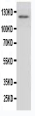
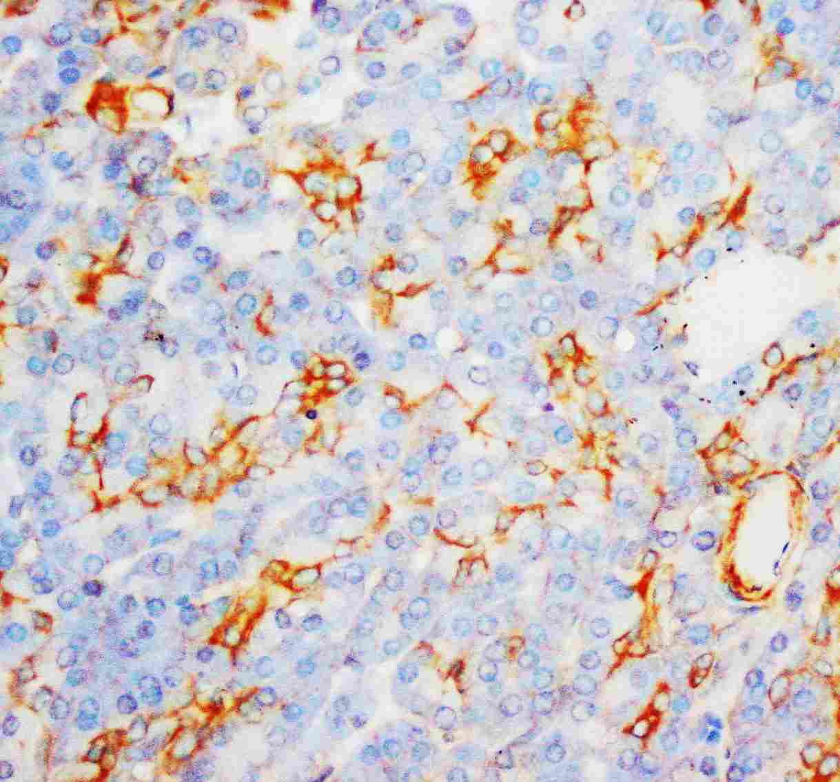
| Description | Rabbit IgG polyclonal antibody for Myeloperoxidase(MPO) detection. Tested with WB, IHC-P in Human;Mouse;Rat. |
| Reconstitution | Add 0.2ml of distilled water will yield a concentration of 500ug/ml. |
| Gene ID | 4353 |
|---|---|
| Other Names | Myeloperoxidase, MPO, 1.11.2.2, Myeloperoxidase, 89 kDa myeloperoxidase, 84 kDa myeloperoxidase, Myeloperoxidase light chain, Myeloperoxidase heavy chain, MPO |
| Calculated MW | 83869 MW KDa |
| Application Details | Immunohistochemistry(Paraffin-embedded Section), 0.5-1 µg/ml, Human, Rat, Mouse, By Heat Western blot, 0.1-0.5 µg/ml, Human, Rat, Mouse |
| Subcellular Localization | Lysosome. |
| Protein Name | Myeloperoxidase(MPO) |
| Contents | Each vial contains 5mg BSA, 0.9mg NaCl, 0.2mg Na2HPO4, 0.05mg Thimerosal, 0.05mg NaN3. |
| Immunogen | A synthetic peptide corresponding to a sequence at the C-terminus of human MPO (714-728aa KNNIFMSNSYPRDFV), different from the related mouse and rat sequences by one amino acid. |
| Purification | Immunogen affinity purified. |
| Cross Reactivity | No cross reactivity with other proteins |
| Storage | At -20˚C for one year. After r˚Constitution, at 4˚C for one month. It˚Can also be aliquotted and stored frozen at -20˚C for a longer time.Avoid repeated freezing and thawing. |
| Sequence Similarities | Belongs to the peroxidase family. XPO subfamily. |
| Name | MPO (HGNC:7218) |
|---|---|
| Function | Part of the host defense system of polymorphonuclear leukocytes. It is responsible for microbicidal activity against a wide range of organisms. In the stimulated PMN, MPO catalyzes the production of hypohalous acids, primarily hypochlorous acid in physiologic situations, and other toxic intermediates that greatly enhance PMN microbicidal activity (PubMed:9922160). Mediates the proteolytic cleavage of alpha-1-microglobulin to form t-alpha-1-microglobulin, which potently inhibits oxidation of low-density lipoprotein particles and limits vascular damage (PubMed:25698971). |
| Cellular Location | Lysosome. |

Thousands of laboratories across the world have published research that depended on the performance of antibodies from Abcepta to advance their research. Check out links to articles that cite our products in major peer-reviewed journals, organized by research category.
info@abcepta.com, and receive a free "I Love Antibodies" mug.

Provided below are standard protocols that you may find useful for product applications.
Background
Myeloperoxidase(MPO) is a mammalian phagocyte hemoprotein though to primarily mediate host defense reactions. It is abundantly expressed in neutrophils and secreted during their activation. Myeloperoxidase is part of the host defense system of human polymorphonuclear leukocytes, responsible for microbicidal activity against a wide range of organisms. It is located in the nucleus as well as in the cytoplasm. Intranuclear MPO may help to protect DNA against damage resulting from oxygen radicals produced during myeloid cell maturation and function.
If you have used an Abcepta product and would like to share how it has performed, please click on the "Submit Review" button and provide the requested information. Our staff will examine and post your review and contact you if needed.
If you have any additional inquiries please email technical services at tech@abcepta.com.
Ordering Information
Other Products
Shipping Information
