


 Foundational characteristics of cancer include proliferation, angiogenesis, migration, evasion of apoptosis, and cellular immortality. Find key markers for these cellular processes and antibodies to detect them.
 The SUMOplot™ Analysis Program predicts and scores sumoylation sites in your protein. SUMOylation is a post-translational modification involved in various cellular processes, such as nuclear-cytosolic transport, transcriptional regulation, apoptosis, protein stability, response to stress, and progression through the cell cycle.
 The Autophagy Receptor Motif Plotter predicts and scores autophagy receptor binding sites in your protein. Identifying proteins connected to this pathway is critical to understanding the role of autophagy in physiological as well as pathological processes such as development, differentiation, neurodegenerative diseases, stress, infection, and cancer.
Anti-Sonic Hedgehog Antibody
- SPECIFICATION
- CITATIONS
- PROTOCOLS
- BACKGROUND

Application
| WB, IHC-P |
|---|---|
| Primary Accession | Q15465 |
| Host | Rabbit |
| Reactivity | Human, Mouse, Rat |
| Clonality | Polyclonal |
| Format | Lyophilized |
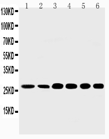
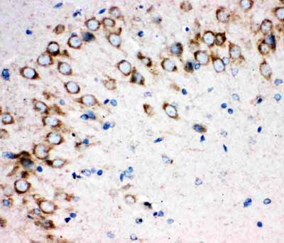
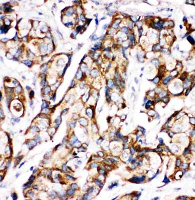
| Description | Rabbit IgG polyclonal antibody for Sonic hedgehog protein(SHH) detection. Tested with WB, IHC-P in Human;Mouse;Rat. |
| Reconstitution | Add 0.2ml of distilled water will yield a concentration of 500ug/ml. |
| Gene ID | 6469 |
|---|---|
| Other Names | Sonic hedgehog protein, SHH, HHG-1, Sonic hedgehog protein N-product, Sonic hedgehog protein C-product, SHH |
| Calculated MW | 49607 MW KDa |
| Application Details | Immunohistochemistry(Paraffin-embedded Section), 0.5-1 µg/ml, Human, Rat, Mouse, By Heat Western blot, 0.1-0.5 µg/ml, Human, Rat, Mouse |
| Subcellular Localization | Sonic hedgehog protein C-product: Secreted, extracellular space . The C-terminal peptide diffuses from the cell. . |
| Tissue Specificity | Expressed in fetal intestine, liver, lung, and kidney. Not expressed in adult tissues. |
| Protein Name | Sonic hedgehog protein |
| Contents | Each vial contains 5mg BSA, 0.9mg NaCl, 0.2mg Na2HPO4, 0.05mg Thimerosal, 0.05mg NaN3. |
| Immunogen | A synthetic peptide corresponding to a sequence at the N-terminus of human Sonic Hedgehog(203-218aa ATVHLEQGGTKLVKDL), identical to the related rat and mouse sequences. |
| Purification | Immunogen affinity purified. |
| Cross Reactivity | No cross reactivity with other proteins |
| Storage | At -20˚C for one year. After r˚Constitution, at 4˚C for one month. It˚Can also be aliquotted and stored frozen at -20˚C for a longer time.Avoid repeated freezing and thawing. |
| Sequence Similarities | Belongs to the hedgehog family. |
| Name | SHH (HGNC:10848) |
|---|---|
| Function | [Sonic hedgehog protein]: The C-terminal part of the sonic hedgehog protein precursor displays an autoproteolysis and a cholesterol transferase activity (By similarity). Both activities result in the cleavage of the full-length protein into two parts (ShhN and ShhC) followed by the covalent attachment of a cholesterol moiety to the C-terminal of the newly generated ShhN (By similarity). Both activities occur in the endoplasmic reticulum (By similarity). Once cleaved, ShhC is degraded in the endoplasmic reticulum (By similarity). |
| Cellular Location | [Sonic hedgehog protein]: Endoplasmic reticulum membrane. Golgi apparatus membrane. Secreted Note=Co-localizes with HHAT in the ER and Golgi membrane |

Thousands of laboratories across the world have published research that depended on the performance of antibodies from Abcepta to advance their research. Check out links to articles that cite our products in major peer-reviewed journals, organized by research category.
info@abcepta.com, and receive a free "I Love Antibodies" mug.

Provided below are standard protocols that you may find useful for product applications.
Background
The mouse, chicken, and zebrafish Shh homologs are highly conserved. SHH expression was not detected in adult tissues examined. However, it was expressed in fetal intestine, liver, lung, and kidney. SHH gene is mapped to 7q. SHH mutations are not a frequent cause of isolated oral clefts.
If you have used an Abcepta product and would like to share how it has performed, please click on the "Submit Review" button and provide the requested information. Our staff will examine and post your review and contact you if needed.
If you have any additional inquiries please email technical services at tech@abcepta.com.
Ordering Information
Other Products
Shipping Information
