


 Foundational characteristics of cancer include proliferation, angiogenesis, migration, evasion of apoptosis, and cellular immortality. Find key markers for these cellular processes and antibodies to detect them.
 The SUMOplot™ Analysis Program predicts and scores sumoylation sites in your protein. SUMOylation is a post-translational modification involved in various cellular processes, such as nuclear-cytosolic transport, transcriptional regulation, apoptosis, protein stability, response to stress, and progression through the cell cycle.
 The Autophagy Receptor Motif Plotter predicts and scores autophagy receptor binding sites in your protein. Identifying proteins connected to this pathway is critical to understanding the role of autophagy in physiological as well as pathological processes such as development, differentiation, neurodegenerative diseases, stress, infection, and cancer.
Anti-VEGFR1/FLT1 Antibody
- SPECIFICATION
- CITATIONS
- PROTOCOLS
- BACKGROUND

Application
| WB |
|---|---|
| Primary Accession | P17948 |
| Host | Rabbit |
| Reactivity | Human |
| Clonality | Polyclonal |
| Format | Lyophilized |
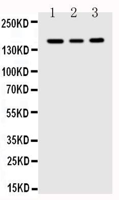
| Description | Rabbit IgG polyclonal antibody for Vascular endothelial growth factor receptor 1(FLT1) detection. Tested with WB in Human. |
| Reconstitution | Add 0.2ml of distilled water will yield a concentration of 500ug/ml. |
| Gene ID | 2321 |
|---|---|
| Other Names | Vascular endothelial growth factor receptor 1, VEGFR-1, 2.7.10.1, Fms-like tyrosine kinase 1, FLT-1, Tyrosine-protein kinase FRT, Tyrosine-protein kinase receptor FLT, FLT, Vascular permeability factor receptor, FLT1, FLT, FRT, VEGFR1 |
| Calculated MW | 150769 MW KDa |
| Application Details | Western blot, 0.1-0.5 µg/ml, Human |
| Subcellular Localization | Isoform 1: Cell membrane; Single-pass type I membrane protein. Endosome. Autophosphorylation promotes ubiquitination and endocytosis. |
| Tissue Specificity | Detected in normal lung, but also in placenta, liver, kidney, heart and brain tissues. Specifically expressed in most of the vascular endothelial cells, and also expressed in peripheral blood monocytes. Isoform 2 is strongly expressed in placenta. Isoform 3 is expressed in corneal epithelial cells (at protein level). Isoform 3 is expressed in vascular smooth muscle cells (VSMC). . |
| Protein Name | Vascular endothelial growth factor receptor 1(VEGFR-1) |
| Contents | Each vial contains 5mg BSA, 0.9mg NaCl, 0.2mg Na2HPO4, 0.05mg Thimerosal, 0.05mg NaN3. |
| Immunogen | A synthetic peptide corresponding to a sequence at the C-terminus of human FLT1 (1299-1317aa HVSEGKRRFTYDHAELERK). |
| Purification | Immunogen affinity purified. |
| Cross Reactivity | No cross reactivity with other proteins |
| Storage | At -20˚C for one year. After r˚Constitution, at 4˚C for one month. It˚Can also be aliquotted and stored frozen at -20˚C for a longer time.Avoid repeated freezing and thawing. |
| Sequence Similarities | Belongs to the protein kinase superfamily. Tyr protein kinase family. CSF-1/PDGF receptor subfamily. |
| Name | FLT1 |
|---|---|
| Synonyms | FLT, FRT, VEGFR1 |
| Function | Tyrosine-protein kinase that acts as a cell-surface receptor for VEGFA, VEGFB and PGF, and plays an essential role in the development of embryonic vasculature, the regulation of angiogenesis, cell survival, cell migration, macrophage function, chemotaxis, and cancer cell invasion. Acts as a positive regulator of postnatal retinal hyaloid vessel regression (By similarity). May play an essential role as a negative regulator of embryonic angiogenesis by inhibiting excessive proliferation of endothelial cells. Can promote endothelial cell proliferation, survival and angiogenesis in adulthood. Its function in promoting cell proliferation seems to be cell-type specific. Promotes PGF-mediated proliferation of endothelial cells, proliferation of some types of cancer cells, but does not promote proliferation of normal fibroblasts (in vitro). Has very high affinity for VEGFA and relatively low protein kinase activity; may function as a negative regulator of VEGFA signaling by limiting the amount of free VEGFA and preventing its binding to KDR. Modulates KDR signaling by forming heterodimers with KDR. Ligand binding leads to the activation of several signaling cascades. Activation of PLCG leads to the production of the cellular signaling molecules diacylglycerol and inositol 1,4,5-trisphosphate and the activation of protein kinase C. Mediates phosphorylation of PIK3R1, the regulatory subunit of phosphatidylinositol 3-kinase, leading to activation of phosphatidylinositol kinase and the downstream signaling pathway. Mediates activation of MAPK1/ERK2, MAPK3/ERK1 and the MAP kinase signaling pathway, as well as of the AKT1 signaling pathway. Phosphorylates SRC and YES1, and may also phosphorylate CBL. Promotes phosphorylation of AKT1 at 'Ser-473'. Promotes phosphorylation of PTK2/FAK1 (PubMed:16685275). |
| Cellular Location | [Isoform 1]: Cell membrane; Single-pass type I membrane protein. Endosome. Note=Autophosphorylation promotes ubiquitination and endocytosis [Isoform 3]: Secreted. [Isoform 5]: Cytoplasm. [Isoform 7]: Cytoplasm. |
| Tissue Location | Detected in normal lung, but also in placenta, liver, kidney, heart and brain tissues. Specifically expressed in most of the vascular endothelial cells, and also expressed in peripheral blood monocytes. Isoform 2 is strongly expressed in placenta. Isoform 3 is expressed in corneal epithelial cells (at protein level). Isoform 3 is expressed in vascular smooth muscle cells (VSMC) |

Thousands of laboratories across the world have published research that depended on the performance of antibodies from Abcepta to advance their research. Check out links to articles that cite our products in major peer-reviewed journals, organized by research category.
info@abcepta.com, and receive a free "I Love Antibodies" mug.

Provided below are standard protocols that you may find useful for product applications.
Background
Vascular endothelial growth factor receptor 1(FLT1) is a protein that in humans is encoded by the FLT1 gene. Oncogene FLT belongs to the src gene family. It is mapped to 13q12. The deduced 1,338-amino acid protein has a calculated molecular mass of 150.6 kD. It has a 758-amino acid extracellular domain, followed by a 22-amino acid transmembrane region and a 558-amino acid cytoplasmic region containing a cluster of basic amino acids and a tyrosine kinase domain. sFLT-1 was identified in placenta, adult lung, kidney, liver and uterus. Like other members of this family, it shows tyrosine protein kinase activity that is important for the control of cell proliferation and differentiation.
If you have used an Abcepta product and would like to share how it has performed, please click on the "Submit Review" button and provide the requested information. Our staff will examine and post your review and contact you if needed.
If you have any additional inquiries please email technical services at tech@abcepta.com.
Ordering Information
Other Products
Shipping Information
