


 Foundational characteristics of cancer include proliferation, angiogenesis, migration, evasion of apoptosis, and cellular immortality. Find key markers for these cellular processes and antibodies to detect them.
 The SUMOplot™ Analysis Program predicts and scores sumoylation sites in your protein. SUMOylation is a post-translational modification involved in various cellular processes, such as nuclear-cytosolic transport, transcriptional regulation, apoptosis, protein stability, response to stress, and progression through the cell cycle.
 The Autophagy Receptor Motif Plotter predicts and scores autophagy receptor binding sites in your protein. Identifying proteins connected to this pathway is critical to understanding the role of autophagy in physiological as well as pathological processes such as development, differentiation, neurodegenerative diseases, stress, infection, and cancer.
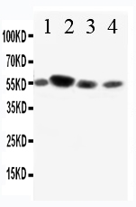
Anti-CD89 Antibody
- SPECIFICATION
- CITATIONS
- PROTOCOLS
- BACKGROUND

Application
| WB |
|---|---|
| Primary Accession | P24071 |
| Host | Rabbit |
| Reactivity | Human |
| Clonality | Polyclonal |
| Format | Lyophilized |
| Description | Rabbit IgG polyclonal antibody for Immunoglobulin alpha Fc receptor(FCAR) detection. Tested with WB in Human. |
| Reconstitution | Add 0.2ml of distilled water will yield a concentration of 500ug/ml. |
| Gene ID | 2204 |
|---|---|
| Other Names | Immunoglobulin alpha Fc receptor, IgA Fc receptor, CD89, FCAR, CD89 |
| Calculated MW | 32265 MW KDa |
| Application Details | Western blot, 0.1-0.5 µg/ml, Human |
| Subcellular Localization | Isoform A.1: Cell membrane; Single-pass type I membrane protein. |
| Tissue Specificity | Isoform A.1, isoform A.2 and isoform A.3 are differentially expressed between blood and mucosal myeloid cells. Isoform A.1, isoform A.2 and isoform A.3 are expressed in monocytes. Isoform A.1 and isoform A.2 are expressed in alveolar macrophages; however only one isoform is expressed at alveolar macrophages surfaces. . |
| Protein Name | Immunoglobulin alpha Fc receptor(IgA Fc receptor) |
| Contents | Each vial contains 5mg BSA, 0.9mg NaCl, 0.2mg Na2HPO4, 0.05mg Thimerosal, 0.05mg NaN3. |
| Immunogen | A synthetic peptide corresponding to a sequence in the middle of human CD89(84-101aa EFVIDHMDANKAGRYQCQ). |
| Purification | Immunogen affinity purified. |
| Cross Reactivity | No cross reactivity with other proteins |
| Storage | At -20˚C for one year. After r˚Constitution, at 4˚C for one month. It˚Can also be aliquotted and stored frozen at -20˚C for a longer time.Avoid repeated freezing and thawing. |
| Name | FCAR |
|---|---|
| Synonyms | CD89 |
| Function | Binds to the Fc region of immunoglobulins alpha. Mediates several functions including cytokine production. |
| Cellular Location | [Isoform A.1]: Cell membrane; Single-pass type I membrane protein [Isoform A.3]: Cell membrane; Single-pass type I membrane protein [Isoform B-delta-S2]: Secreted. |
| Tissue Location | Isoform A.1, isoform A.2 and isoform A.3 are differentially expressed between blood and mucosal myeloid cells Isoform A.1, isoform A.2 and isoform A.3 are expressed in monocytes Isoform A.1 and isoform A.2 are expressed in alveolar macrophages; however only one isoform is expressed at alveolar macrophages surfaces |

Thousands of laboratories across the world have published research that depended on the performance of antibodies from Abcepta to advance their research. Check out links to articles that cite our products in major peer-reviewed journals, organized by research category.
info@abcepta.com, and receive a free "I Love Antibodies" mug.

Provided below are standard protocols that you may find useful for product applications.
Background
FCAR, Receptor for Fc fragment of IGA, is also known as CD89. Human Fc-alpha receptor(FCAR) is present on a number of cell types, including neutrophils, monocytes, macrophages, and eosinophils. FCAR interacts with aggregated IgAs, such as IgA coated on the surface of an invading microorganism, and mediates several immunologic defense processes such as phagocytosis, antibody-dependent cell-mediated cytotoxicity, and stimulation of the release of inflammatory mediators. FCAR is a glycoprotein of 50 to 100 kD, with diversity on different cell types. FCAR is mapped to 19q13.4. Human COS cells transfected with FCAR cDNA bind to IgA, but not IgG.
If you have used an Abcepta product and would like to share how it has performed, please click on the "Submit Review" button and provide the requested information. Our staff will examine and post your review and contact you if needed.
If you have any additional inquiries please email technical services at tech@abcepta.com.
Ordering Information
Other Products
Shipping Information
