


 Foundational characteristics of cancer include proliferation, angiogenesis, migration, evasion of apoptosis, and cellular immortality. Find key markers for these cellular processes and antibodies to detect them.
 The SUMOplot™ Analysis Program predicts and scores sumoylation sites in your protein. SUMOylation is a post-translational modification involved in various cellular processes, such as nuclear-cytosolic transport, transcriptional regulation, apoptosis, protein stability, response to stress, and progression through the cell cycle.
 The Autophagy Receptor Motif Plotter predicts and scores autophagy receptor binding sites in your protein. Identifying proteins connected to this pathway is critical to understanding the role of autophagy in physiological as well as pathological processes such as development, differentiation, neurodegenerative diseases, stress, infection, and cancer.
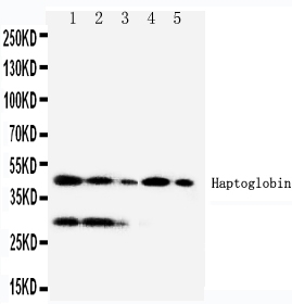
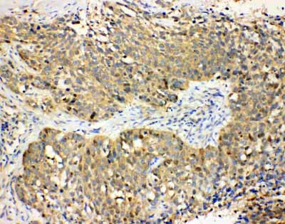
Anti-Haptoglobin Antibody
- SPECIFICATION
- CITATIONS
- PROTOCOLS
- BACKGROUND

Application
| WB, IHC-P |
|---|---|
| Primary Accession | P00738 |
| Host | Rabbit |
| Reactivity | Human |
| Clonality | Polyclonal |
| Format | Lyophilized |
| Description | Rabbit IgG polyclonal antibody for Haptoglobin(HP) detection. Tested with WB, IHC-P in Human. |
| Reconstitution | Add 0.2ml of distilled water will yield a concentration of 500ug/ml. |
| Gene ID | 3240 |
|---|---|
| Other Names | Haptoglobin, Zonulin, Haptoglobin alpha chain, Haptoglobin beta chain, HP |
| Calculated MW | 45205 MW KDa |
| Application Details | Immunohistochemistry(Paraffin-embedded Section), 0.5-1 µg/ml, Human, By Heat Western blot, 0.1-0.5 µg/ml, Human |
| Subcellular Localization | Secreted. |
| Tissue Specificity | Expressed by the liver and secreted in plasma. |
| Protein Name | Haptoglobin |
| Contents | Each vial contains 5mg BSA, 0.9mg NaCl, 0.2mg Na2HPO4, 0.05mg Thimerosal, 0.05mg NaN3. |
| Immunogen | A synthetic peptide corresponding to a sequence in the middle region of human Haptoglobin(294-309aa DHLKYVMLPVADQDQC). |
| Purification | Immunogen affinity purified. |
| Cross Reactivity | No cross reactivity with other proteins |
| Storage | At -20˚C for one year. After r˚Constitution, at 4˚C for one month. It˚Can also be aliquotted and stored frozen at -20˚C for a longer time.Avoid repeated freezing and thawing. |
| Sequence Similarities | Belongs to the peptidase S1 family. |
| Name | HP |
|---|---|
| Function | As a result of hemolysis, hemoglobin is found to accumulate in the kidney and is secreted in the urine. Haptoglobin captures, and combines with free plasma hemoglobin to allow hepatic recycling of heme iron and to prevent kidney damage. Haptoglobin also acts as an antioxidant, has antibacterial activity, and plays a role in modulating many aspects of the acute phase response. Hemoglobin/haptoglobin complexes are rapidly cleared by the macrophage CD163 scavenger receptor expressed on the surface of liver Kupfer cells through an endocytic lysosomal degradation pathway. |
| Cellular Location | Secreted. |
| Tissue Location | Expressed by the liver and secreted in plasma. |

Thousands of laboratories across the world have published research that depended on the performance of antibodies from Abcepta to advance their research. Check out links to articles that cite our products in major peer-reviewed journals, organized by research category.
info@abcepta.com, and receive a free "I Love Antibodies" mug.

Provided below are standard protocols that you may find useful for product applications.
Background
Haptoglobin(HP), is a protein that in humans is encoded by the HP gene. Haptoglobin, a plasma glycoprotein that binds free hemoglobin, has a tetrameric structure of 2 alpha and 2 beta polypeptides that are covalently associated by disulfide bonds. Haptoglobin is homologous to serine proteases of the chymotrypsinogen family. In the chimpanzee, there are 3 genes in the haptoglobin family(haptoglobin, HP; haptoglobin-related, HPR; and haptoglobin-primate, HPP), whereas only 2 genes exist in humans(HP and HPR). There are similarities between the primary structures of the alpha chain and of light chains of gamma globulins. The alpha haptoglobin locus is on the long arm of chromosome 16, where its exact cytogenetic location is 16q22.2. A major function of haptoglobin is to bind hemoglobin(Hb) to form a stable Hp-Hb complex and thereby prevent Hb-induced oxidative tissue damage. Haptoglobin is an unusual secretory protein in that it is proteolytically processed in the endoplasmic reticulum and not in the Golgi. The human haptoglobin HP*2 allele contains a 1.7-kb intragenic duplication that arose after a unique nonhomologous recombination between the prototype HP*1 alleles.
If you have used an Abcepta product and would like to share how it has performed, please click on the "Submit Review" button and provide the requested information. Our staff will examine and post your review and contact you if needed.
If you have any additional inquiries please email technical services at tech@abcepta.com.
Ordering Information
Other Products
Shipping Information
