


 Foundational characteristics of cancer include proliferation, angiogenesis, migration, evasion of apoptosis, and cellular immortality. Find key markers for these cellular processes and antibodies to detect them.
 The SUMOplot™ Analysis Program predicts and scores sumoylation sites in your protein. SUMOylation is a post-translational modification involved in various cellular processes, such as nuclear-cytosolic transport, transcriptional regulation, apoptosis, protein stability, response to stress, and progression through the cell cycle.
 The Autophagy Receptor Motif Plotter predicts and scores autophagy receptor binding sites in your protein. Identifying proteins connected to this pathway is critical to understanding the role of autophagy in physiological as well as pathological processes such as development, differentiation, neurodegenerative diseases, stress, infection, and cancer.
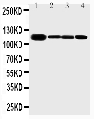
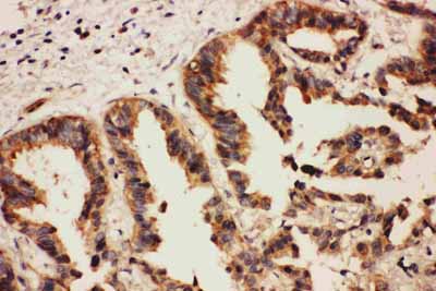
Anti-Eph Receptor A2 Antibody
- SPECIFICATION
- CITATIONS
- PROTOCOLS
- BACKGROUND

Application
| WB, IHC-P |
|---|---|
| Primary Accession | P29317 |
| Host | Rabbit |
| Reactivity | Human, Mouse, Rat |
| Clonality | Polyclonal |
| Format | Lyophilized |
| Description | Rabbit IgG polyclonal antibody for Ephrin type-A receptor 2(EPHA2) detection. Tested with WB, IHC-P in Human;Mouse;Rat. |
| Reconstitution | Add 0.2ml of distilled water will yield a concentration of 500ug/ml. |
| Gene ID | 1969 |
|---|---|
| Other Names | Ephrin type-A receptor 2, 2.7.10.1, Epithelial cell kinase, Tyrosine-protein kinase receptor ECK, EPHA2, ECK |
| Calculated MW | 108266 MW KDa |
| Application Details | Immunohistochemistry(Paraffin-embedded Section), 0.5-1 µg/ml, Human, Mouse, Rat, By Heat Western blot, 0.1-0.5 µg/ml, Human, Rat, Mouse |
| Subcellular Localization | Cell membrane; Single-pass type I membrane protein. Cell projection, ruffle membrane; Single-pass type I membrane protein. Cell projection, lamellipodium membrane; Single- pass type I membrane protein. Cell junction, focal adhesion. Present at regions of cell-cell contacts but also at the leading edge of migrating cells. |
| Tissue Specificity | Expressed in brain and glioma tissue and glioma cell lines (at protein level). Expressed most highly in tissues that contain a high proportion of epithelial cells, e.g. skin, intestine, lung, and ovary. . |
| Protein Name | Ephrin type-A receptor 2 |
| Contents | Each vial contains 5mg BSA, 0.9mg NaCl, 0.2mg Na2HPO4, 0.05mg Thimerosal, 0.05mg NaN3. |
| Immunogen | A synthetic peptide corresponding to a sequence at the C-terminus of human Eph receptor A2(954-976aa HQKRIAYSLLGLKDQVNTVGIPI), identical to the related rat and mouse sequences. |
| Purification | Immunogen affinity purified. |
| Cross Reactivity | No cross reactivity with other proteins |
| Storage | At -20˚C for one year. After r˚Constitution, at 4˚C for one month. It˚Can also be aliquotted and stored frozen at -20˚C for a longer time.Avoid repeated freezing and thawing. |
| Sequence Similarities | Belongs to the protein kinase superfamily. Tyr protein kinase family. Ephrin receptor subfamily. |
| Name | EPHA2 |
|---|---|
| Synonyms | ECK |
| Function | Receptor tyrosine kinase which binds promiscuously membrane- bound ephrin-A family ligands residing on adjacent cells, leading to contact-dependent bidirectional signaling into neighboring cells. The signaling pathway downstream of the receptor is referred to as forward signaling while the signaling pathway downstream of the ephrin ligand is referred to as reverse signaling. Activated by the ligand ephrin- A1/EFNA1 regulates migration, integrin-mediated adhesion, proliferation and differentiation of cells. Regulates cell adhesion and differentiation through DSG1/desmoglein-1 and inhibition of the ERK1/ERK2 (MAPK3/MAPK1, respectively) signaling pathway. May also participate in UV radiation-induced apoptosis and have a ligand- independent stimulatory effect on chemotactic cell migration. During development, may function in distinctive aspects of pattern formation and subsequently in development of several fetal tissues. Involved for instance in angiogenesis, in early hindbrain development and epithelial proliferation and branching morphogenesis during mammary gland development. Engaged by the ligand ephrin-A5/EFNA5 may regulate lens fiber cells shape and interactions and be important for lens transparency development and maintenance. With ephrin-A2/EFNA2 may play a role in bone remodeling through regulation of osteoclastogenesis and osteoblastogenesis. |
| Cellular Location | Cell membrane; Single-pass type I membrane protein. Cell projection, ruffle membrane; Single-pass type I membrane protein. Cell projection, lamellipodium membrane; Single-pass type I membrane protein. Cell junction, focal adhesion. Note=Present at regions of cell-cell contacts but also at the leading edge of migrating cells (PubMed:19573808, PubMed:20861311). Relocates from the plasma membrane to the cytoplasmic and perinuclear regions in cancer cells (PubMed:18794797). |
| Tissue Location | Expressed in brain and glioma tissue and glioma cell lines (at protein level). Expressed most highly in tissues that contain a high proportion of epithelial cells, e.g. skin, intestine, lung, and ovary. |

Thousands of laboratories across the world have published research that depended on the performance of antibodies from Abcepta to advance their research. Check out links to articles that cite our products in major peer-reviewed journals, organized by research category.
info@abcepta.com, and receive a free "I Love Antibodies" mug.

Provided below are standard protocols that you may find useful for product applications.
Background
EPHA2(ephrin type-A receptor 2) also known as ECK, is a protein that in humans is encoded by the EPHA2 gene. This gene belongs to the ephrin receptor subfamily of the protein-tyrosine kinase family. Receptors in the EPH subfamily typically have a single kinase domain and an extracellular region containing a Cys-rich domain and 2 fibronectin type III repeats. By somatic cell hybrid analysis and fluorescence in situ hybridization, the EPHA2 gene is mapped to chromosome 1p36.1. By screening a HeLa cell cDNA library with degenerate oligonucleotides based on highly conserved regions of receptor protein-tyrosine kinases, Lindberg and Hunter isolated cDNAs encoding EPHA2, which they called ECK. EPHA2 was readily detectable in human lens fiber cells using immunoblot and immunohistochemistry. EGFR and EPHA2 mediated HCV entry by regulating CD81 -claudin-1(CLDN1) coreceptor associations and viral glycoprotein-dependent membrane fusion.
If you have used an Abcepta product and would like to share how it has performed, please click on the "Submit Review" button and provide the requested information. Our staff will examine and post your review and contact you if needed.
If you have any additional inquiries please email technical services at tech@abcepta.com.
Ordering Information
Other Products
Shipping Information
